Biotech & biologics lab testing
Quality control of therapeutic proteins and other biologics: content, purity, variants and process impurities. 9 test methods and 19 instruments, explained step by step.
- ICH Q6B
- ICH Q5
- USP
- Ph. Eur.
- WHO
Biologic drug substance & product
Protein content, purity, size and charge variants, identity.
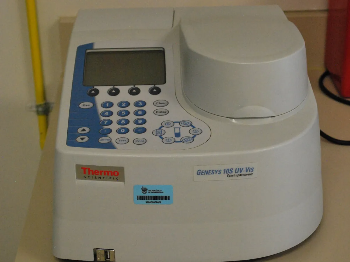
- UV spectrophotometry
Protein Concentration by UV Absorbance at 280 nm (A280)
The protein solution is diluted with its formulation buffer so the absorbance is in the linear range, absorbance is measured at 280 nm (with a 320 nm scatter correction) against the buffer blank, and concentration is calculated with the Beer–Lambert law using the protein's extinction coefficient.
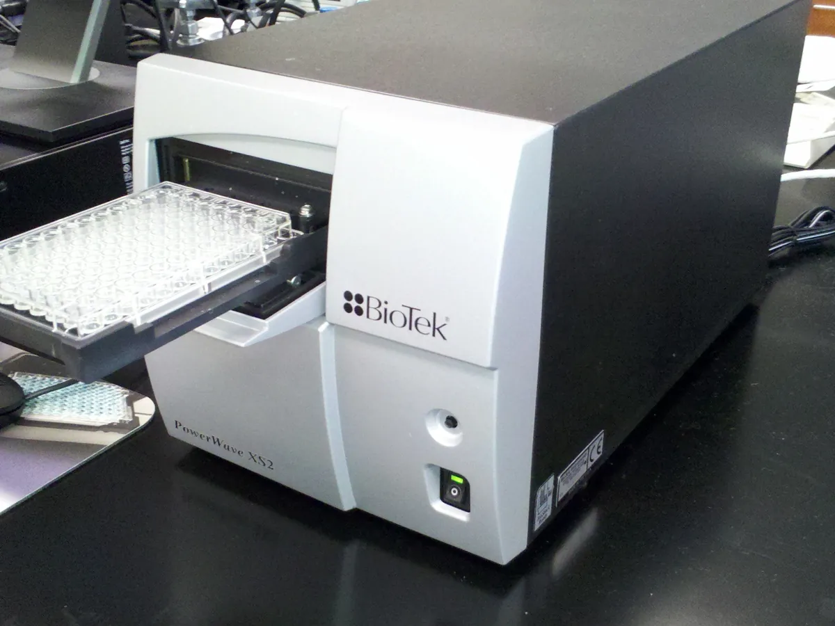
Open method - Microplate colorimetry
Total Protein by BCA or Bradford Microplate Assay
Samples and a series of protein standards react with a colour reagent in a 96-well plate. Absorbance is read on a microplate reader (562 nm for BCA, 595 nm for Bradford), a standard curve is fitted, and sample protein concentration is read from the curve and multiplied by the dilution factor.
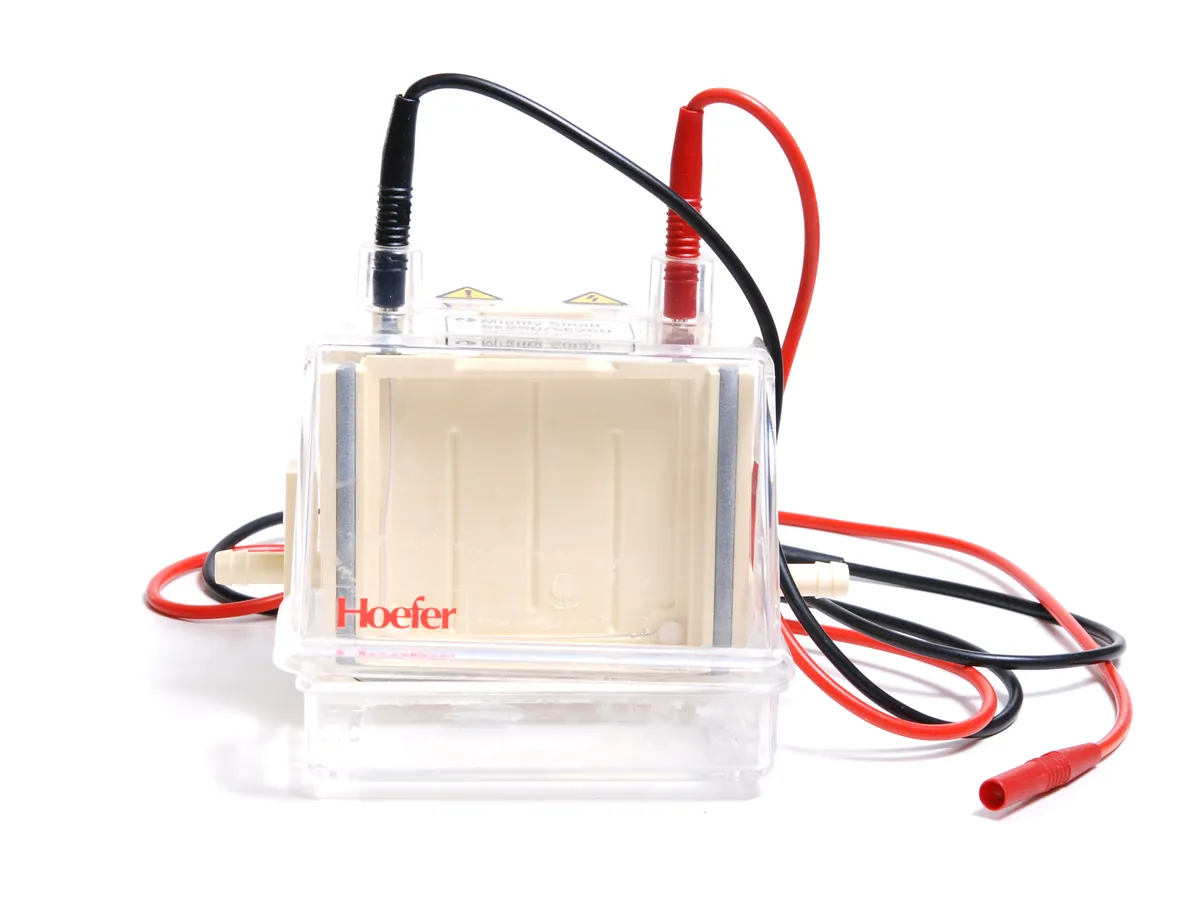
Open method - SDS polyacrylamide gel electrophoresis with Coomassie staining and densitometry
Purity by SDS-PAGE (reduced and non-reduced)
Separates the protein and its fragments, aggregates and other protein impurities by size on a polyacrylamide gel. The stained bands are scanned and the main band (or bands) is expressed as a percentage of all the protein in the lane. Running the sample both reduced and non-reduced shows different kinds of impurity.
Open method - Size-exclusion HPLC with UV detection (isocratic, non-denaturing)
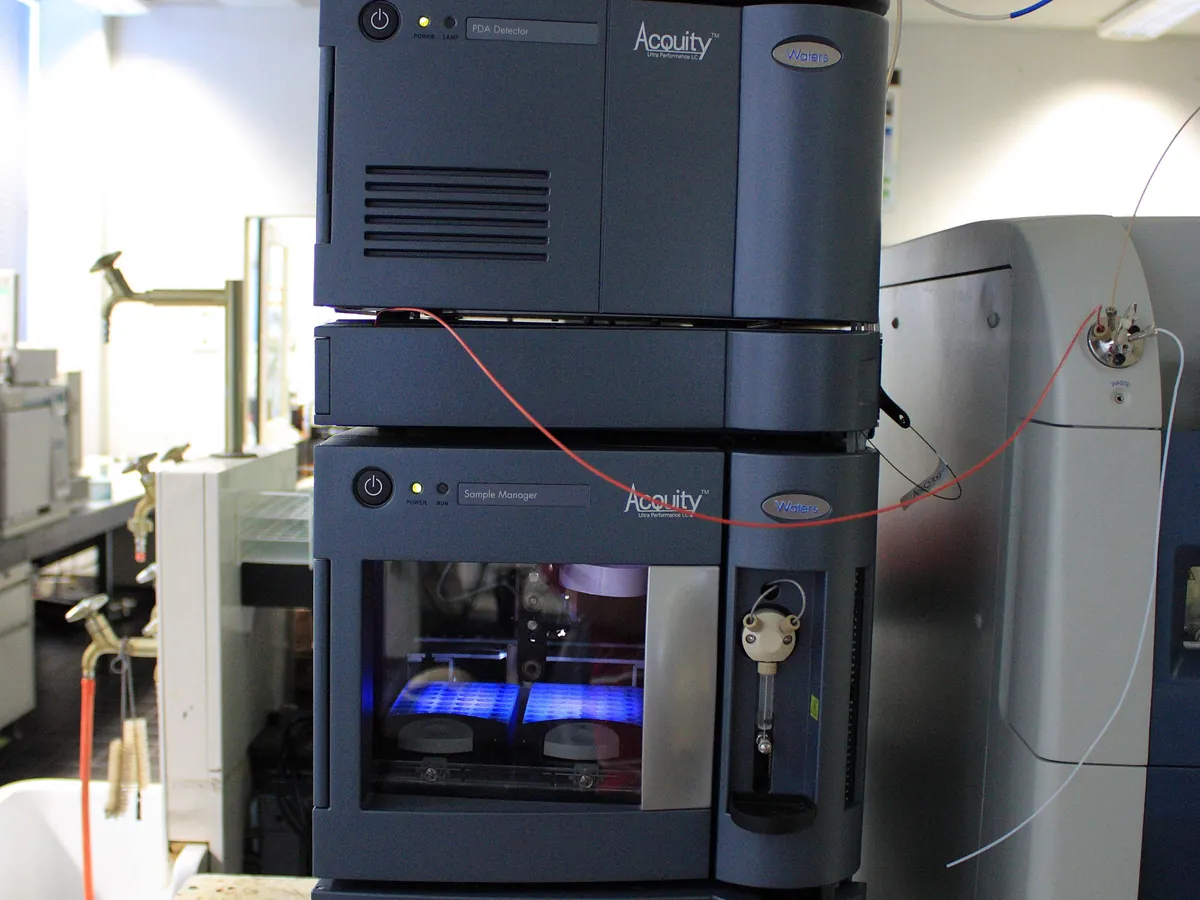
Aggregates by size-exclusion HPLC (SEC-HPLC)
Separates the protein by molecular size under native conditions. Aggregates (dimers and larger) elute first, then the monomer, then fragments. The result is reported as % monomer, % HMW and % LMW by area normalisation. Aggregate level is a critical quality attribute because aggregates can reduce activity and increase the risk of an immune response.
Open method - Capillary isoelectric focusing with UV (280 nm) whole-column imaging or mobilisation
Charge variants by capillary isoelectric focusing (cIEF / icIEF)
Separates the protein's charge variants by their isoelectric point (pI) in a pH gradient inside a capillary. Peaks are assigned to acidic, main and basic regions and reported as area percentages. Two pI markers in every sample let the pI of each peak be calculated.
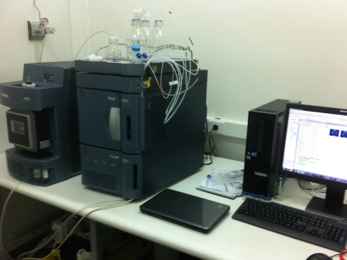
Open method - Enzymatic digestion, reversed-phase UHPLC with UV and high-resolution MS/MS
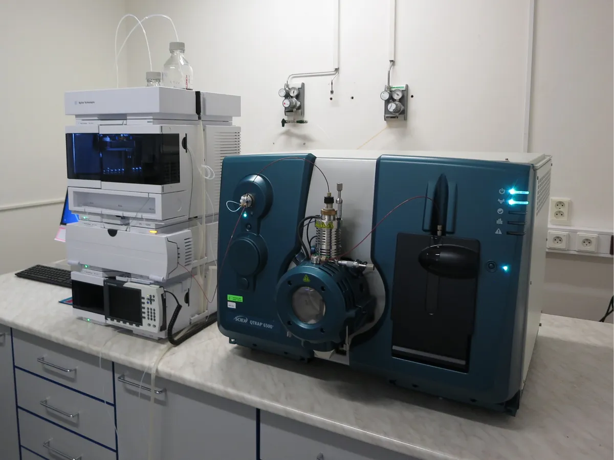
Identity by peptide mapping (enzymatic digest with LC-MS)
The protein is denatured, reduced, alkylated and cut into peptides with trypsin. The peptides are separated by reversed-phase UHPLC and detected by UV and mass spectrometry. The peptide pattern is compared with the reference standard to confirm identity, and the MS data confirm which parts of the sequence were seen (sequence coverage) and pick up modifications such as oxidation or deamidation.
Open method
Process-related impurities
Host cell protein and residual DNA.
- Sandwich ELISA (polyclonal anti-HCP antibodies, colorimetric read-out)
Host cell protein (HCP) by sandwich ELISA
Measures the total amount of protein from the host cell line that remains in the purified product. A polyclonal antibody mix captures many different HCPs at once; the signal is read against a standard curve of the HCP standard and reported as ng HCP per mg of product (ppm). Dilution linearity and spike recovery show the product itself does not distort the result.
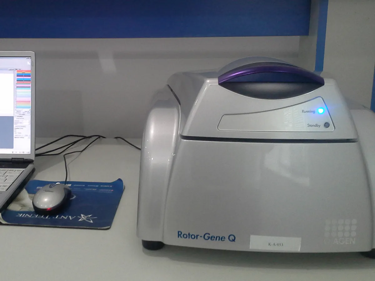
Open method - DNA extraction followed by real-time quantitative PCR against a genomic DNA standard curve
Residual host cell DNA by quantitative PCR (qPCR)
Measures the very small amount of DNA from the host cells left in the purified protein. DNA is extracted from the sample, a sequence that occurs many times in the host genome is amplified by qPCR, and the amount is read from a standard curve of host-cell genomic DNA. The result is reported per mg of product and per dose; a commonly applied limit is 10 ng per dose (typical, from WHO guidance).
Open method
Cells & bioprocess
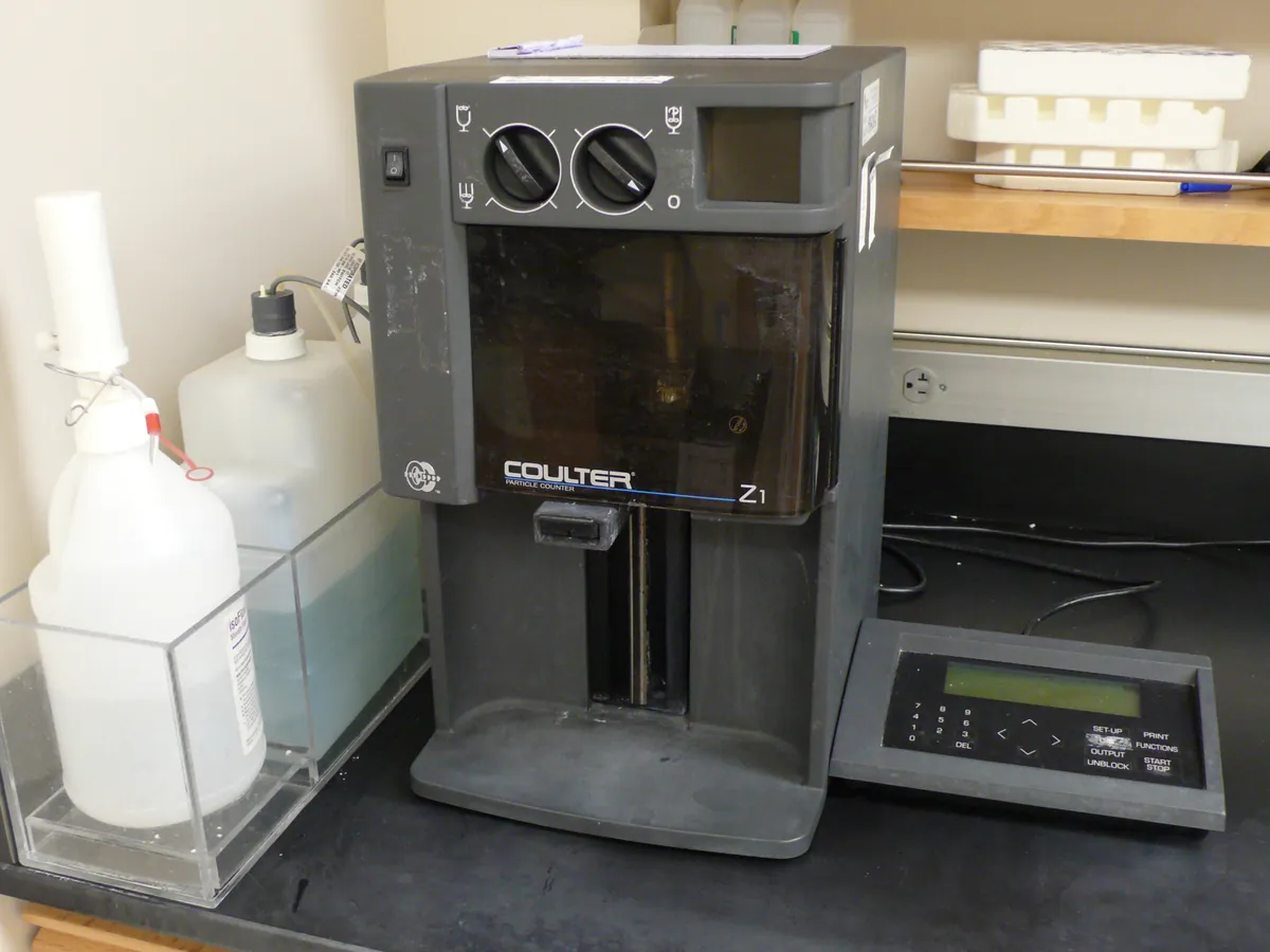
Cell counts and viability in culture.
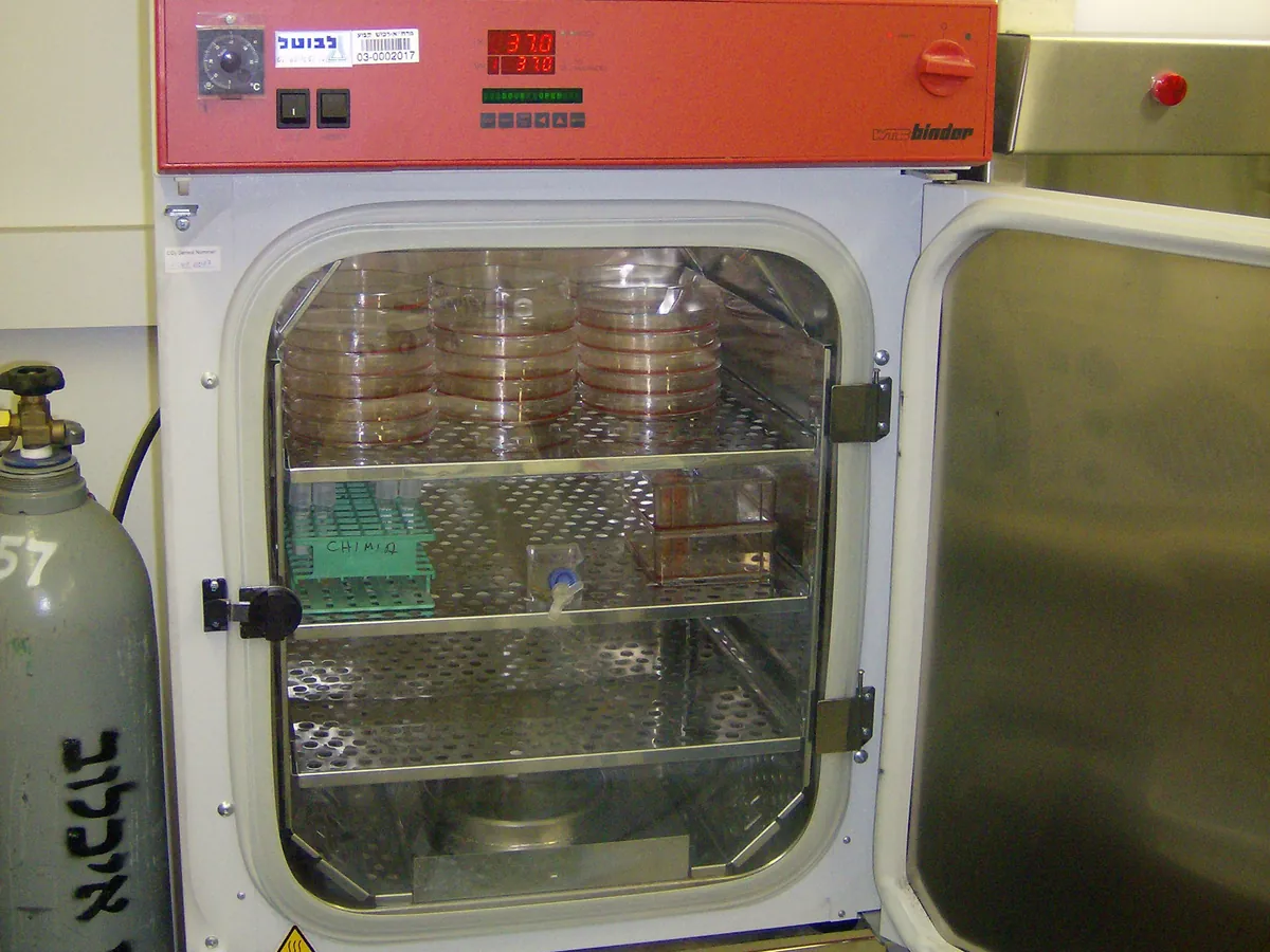
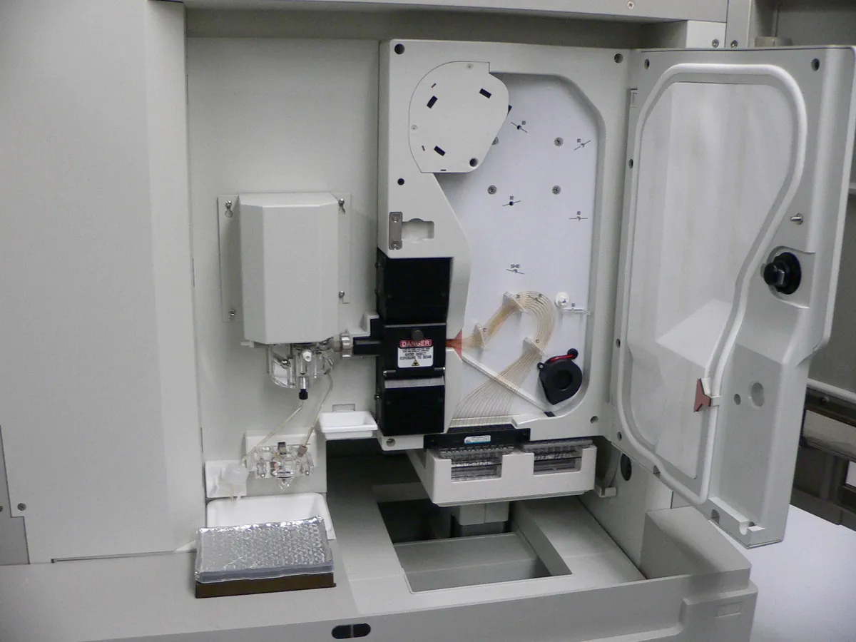
Instruments in a biotech & biologics lab
Learning paths
Guides
- QC of biologics: what is tested and whyA plain-language map of quality control for therapeutic proteins such as monoclonal antibodies: the ICH Q6B attribute groups (identity, purity and impurities, potency, quantity), the difference between product-related and process-related impurities, and how comparability and stability testing fit in.
- Analytical method validation (ICH Q2(R2))What method validation is, how it differs from verification and transfer, the validation characteristics with typical acceptance criteria for assay and impurity methods, and how a validation runs from protocol to report.