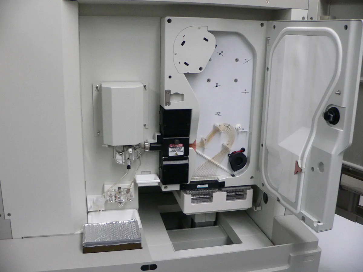
Capillary electrophoresis system
Separates charged molecules in a thin fused-silica capillary under a high voltage (typically up to 30 kV). Used in biologics QC for size purity (CE-SDS), charge variants (cIEF, CZE) and glycan analysis.
- Biotechnology
- Biopharmaceutical
- Pharmaceutical
- Genomics
- Forensic science
- Academic research
What is CE?
Capillary electrophoresis (CE) does the same job as a slab gel, but inside a fused-silica capillary about 25–100 µm wide and 30–100 cm long. The narrow capillary loses heat quickly, so very high voltages can be used. Separations are fast and sharp, and an on-capillary UV or fluorescence detector gives quantitative peaks like a chromatogram.
Several CE modes are used for proteins. CE-SDS fills the capillary with a sieving gel and separates SDS-coated proteins by size; it has largely replaced SDS-PAGE for quantitative purity of monoclonal antibodies. Capillary isoelectric focusing (cIEF) separates charge variants by their isoelectric point. Capillary zone electrophoresis (CZE) separates by charge-to-size ratio in free solution.
Temperature and voltage control are critical: both change migration times and resolution. CE systems therefore have a thermostatted capillary cartridge and a precisely regulated high-voltage supply, and both are verified during qualification and routine calibration.
Used for
- Size purity of monoclonal antibodies by CE-SDS (reduced and non-reduced)
- Charge variants by cIEF or CZE
- Released N-glycan profiling with fluorescent labels (CE-LIF)
- Peptide and small-molecule separations, chiral separations
- DNA fragment sizing and oligonucleotide purity (capillary gel electrophoresis)
How it works
- 01
Condition
The capillary is rinsed with conditioning solutions and filled with background electrolyte or sieving gel.
- 02
Inject
A small plug of sample is introduced by pressure or by voltage (electrokinetic injection).
- 03
Apply voltage
High voltage is applied across the capillary ends dipping in buffer vials.
- 04
Separate
Molecules migrate at different speeds towards the detector window.
- 05
Detect
UV (often 214 or 220 nm, 280 nm for cIEF) or laser-induced fluorescence detects analytes through a clear window in the capillary.
- 06
Integrate
Software integrates peaks and calculates corrected peak areas and relative per cent.
Advantages
- High resolution and quantitative peaks
- Very small sample volumes (nanolitres injected)
- Low reagent use and little organic solvent
- Automated runs of many samples
- Several separation modes on one instrument
Limitations
- Lower concentration sensitivity with UV because of the short path length
- Migration times can shift with capillary surface changes
- Injection precision is lower than HPLC unless an internal standard is used
- Capillaries and gel buffers need careful conditioning
- High voltage requires interlocks and careful handling
1 calibration procedure
Methods that use CE
Keep every CE in control: usage logs, calibration and maintenance schedules, and automatic blocking of overdue instruments.