Identity by peptide mapping (enzymatic digest with LC-MS)
The protein is denatured, reduced, alkylated and cut into peptides with trypsin. The peptides are separated by reversed-phase UHPLC and detected by UV and mass spectrometry. The peptide pattern is compared with the reference standard to confirm identity, and the MS data confirm which parts of the sequence were seen (sequence coverage) and pick up modifications such as oxidation or deamidation.
- Biotech & biologics
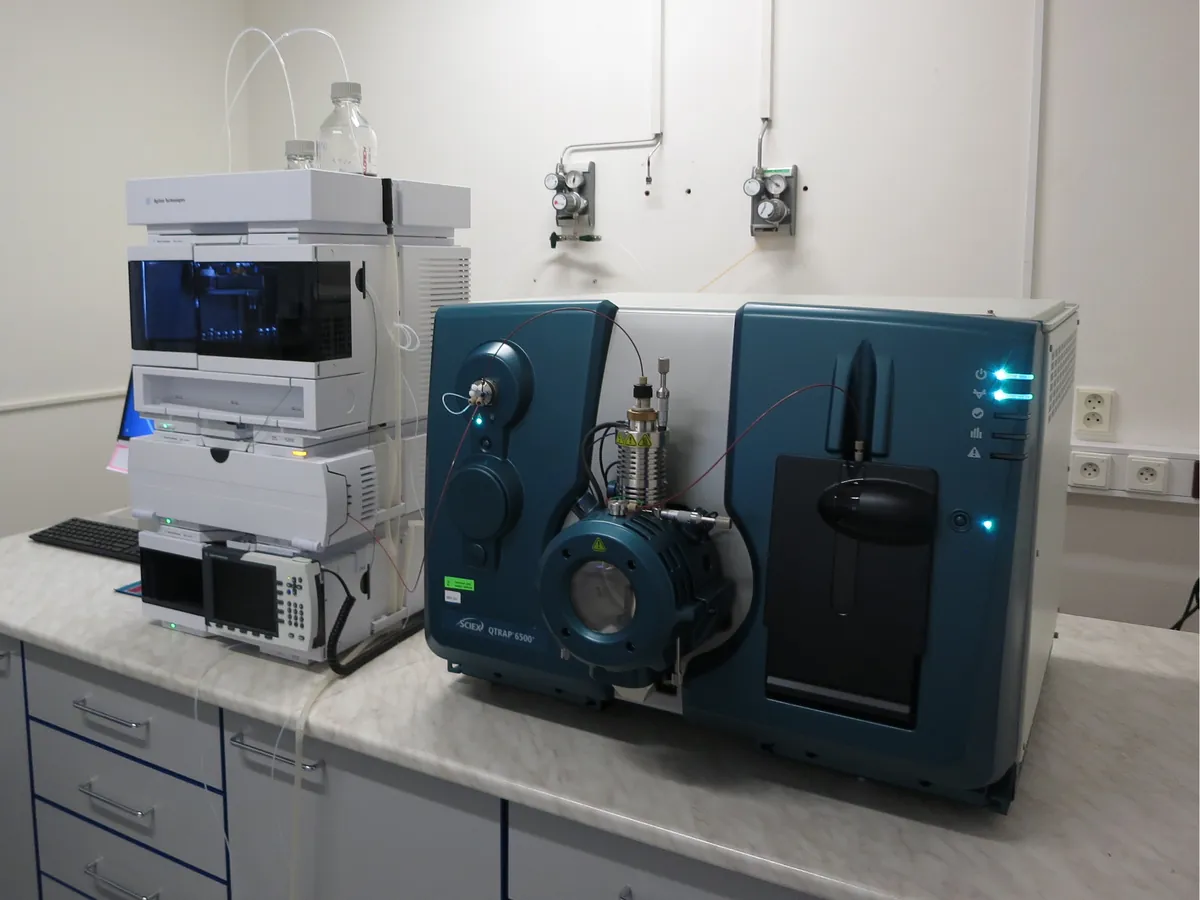
- Enzymatic digestion, reversed-phase UHPLC with UV and high-resolution MS/MS
- LC-MS
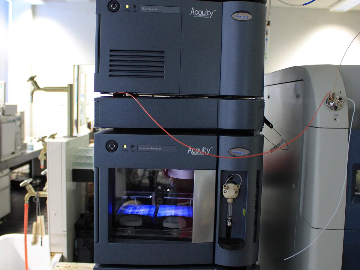
- UHPLC
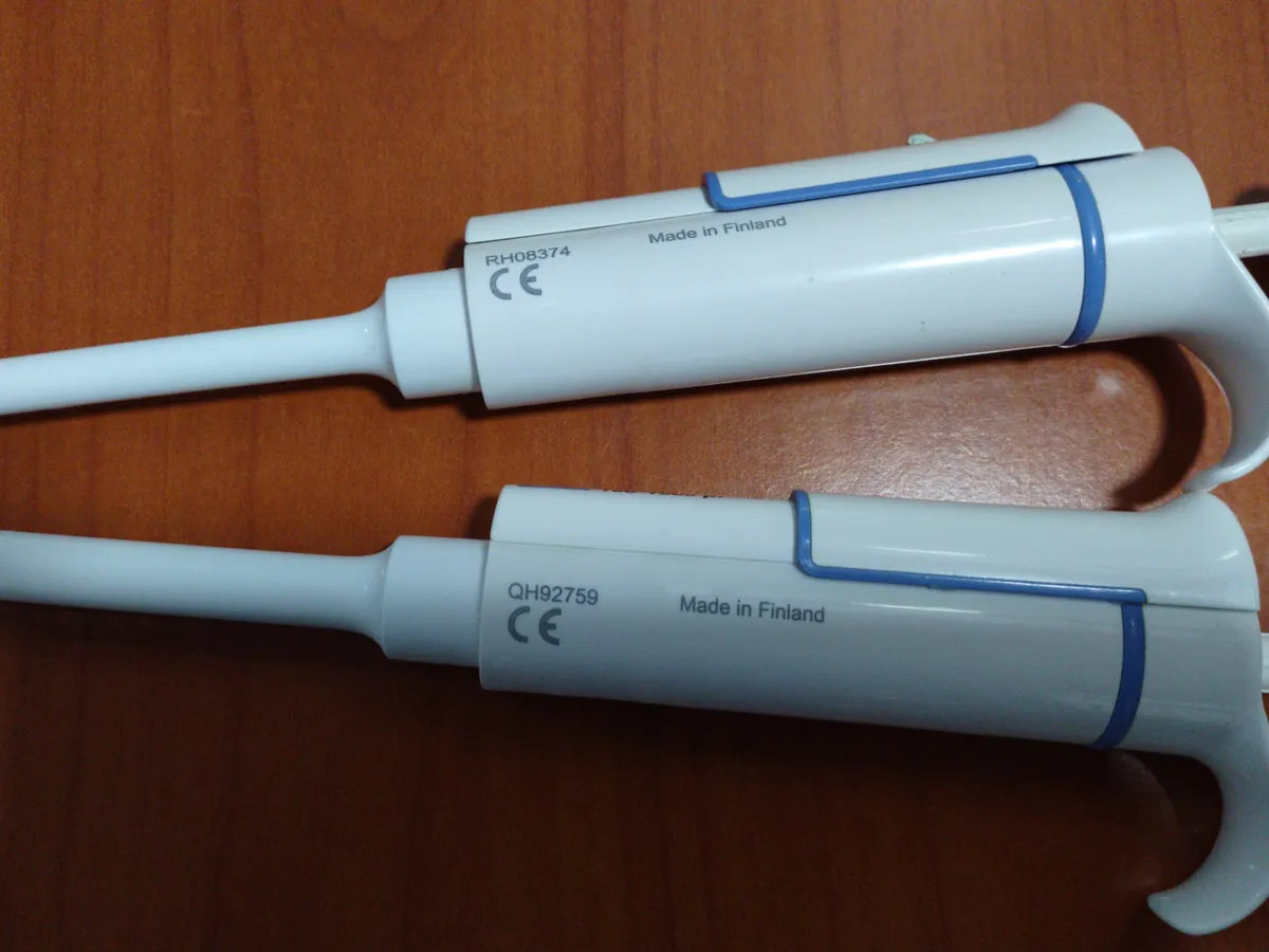
- Micropipette
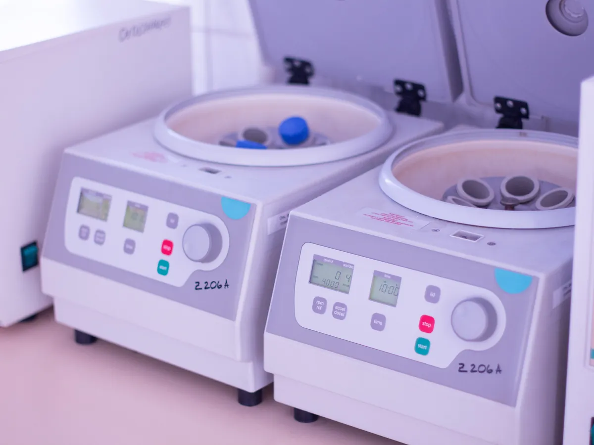
- Centrifuge
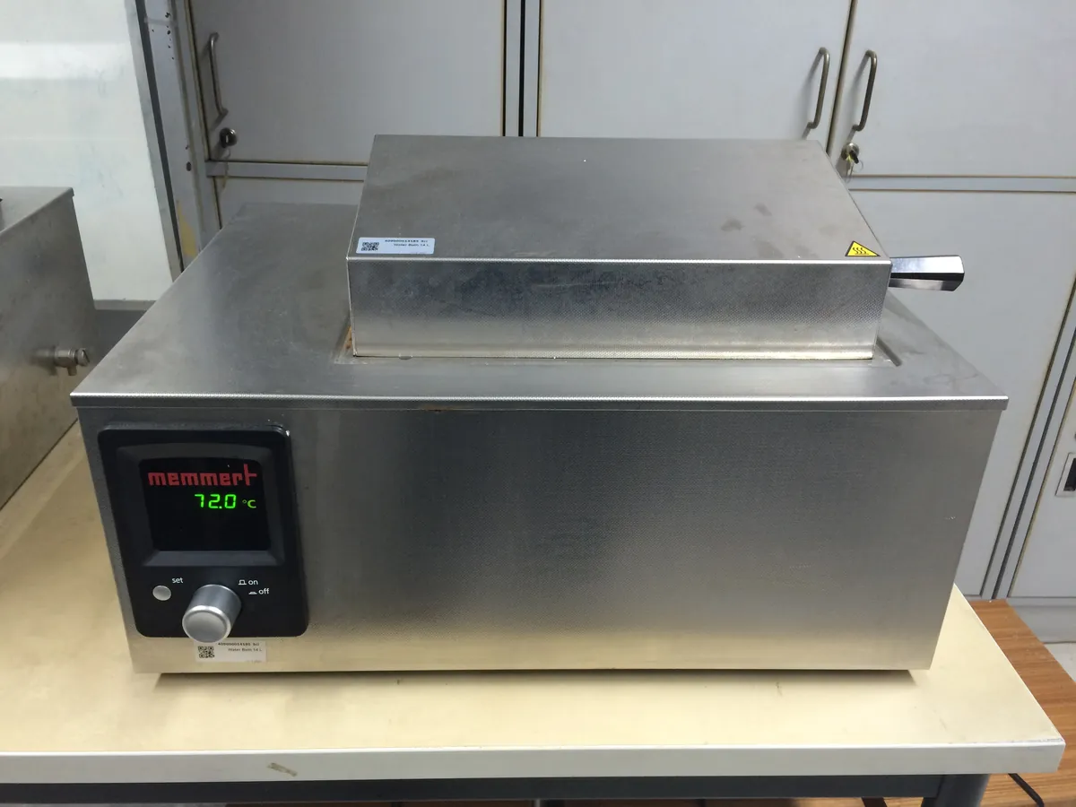
- Water bath
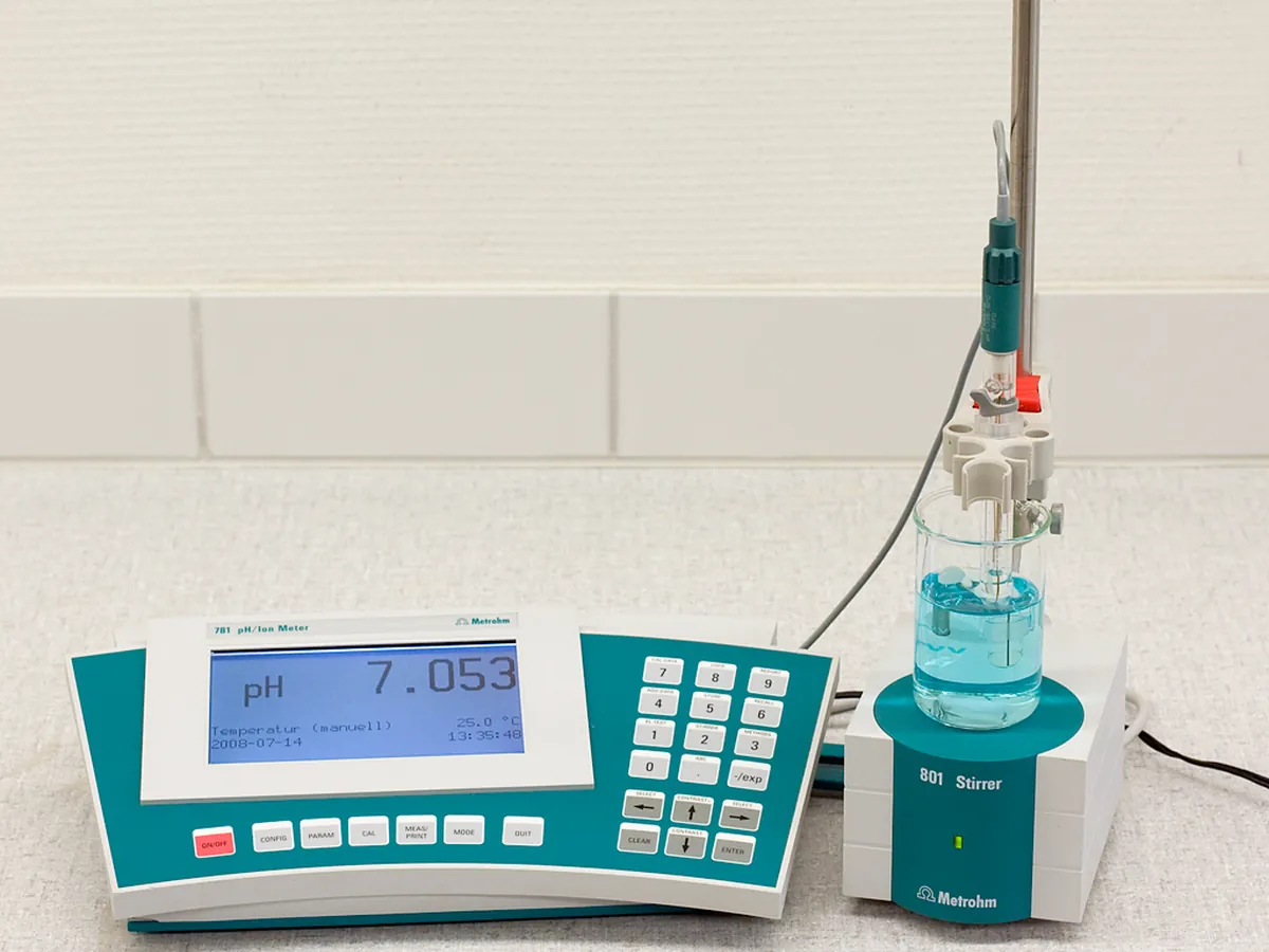
- pH meter
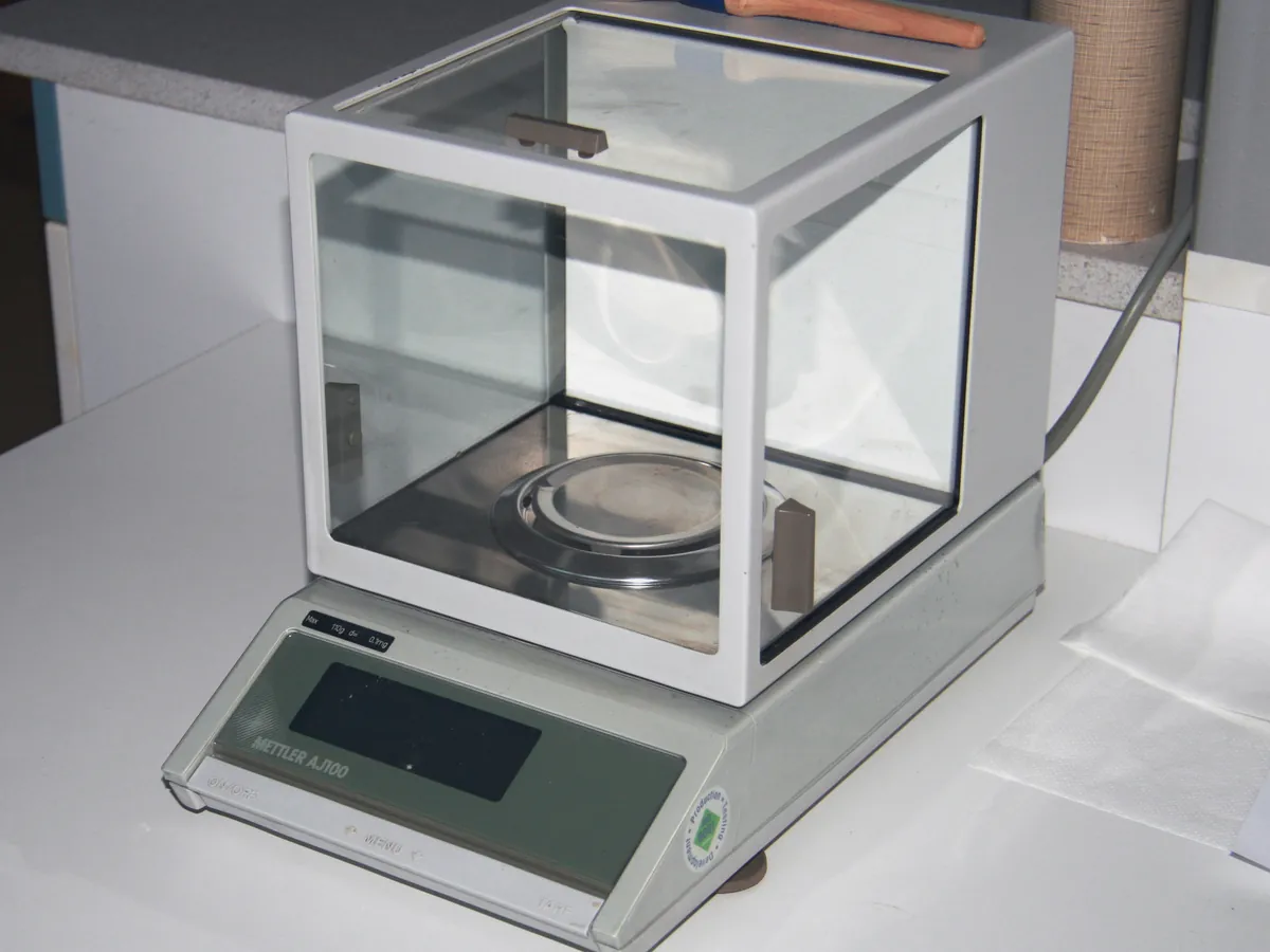
- Analytical balance
Reference: Typical method based on Ph. Eur. 2.2.55, USP <1055> and ICH Q6B. Follow your approved test procedure and product specification.
Why this method works
A protein is too big to compare residue by residue in one measurement, so it is cut at predictable places. Trypsin cleaves after lysine and arginine (not before proline), so a given sequence always gives the same set of peptides — a 'fingerprint'. Before digestion, the protein is unfolded with a denaturant (guanidine hydrochloride or urea), disulphide bonds are reduced with DTT and the free cysteines are capped with iodoacetamide so they cannot re-form bonds. Reversed-phase chromatography separates the peptides by hydrophobicity with a water–acetonitrile gradient containing a small amount of acid. UV at 214 nm gives the familiar map profile. Electrospray mass spectrometry measures each peptide's mass very accurately, and MS/MS breaks it into fragment ions that confirm its sequence. Matching observed peptides to the theoretical digest gives the sequence coverage and confirms the identity of the molecule; a single amino acid change shifts the mass of one peptide and is detected.
Instruments used
Reagents & standards
| Reagent | Grade / specification |
|---|---|
| Denaturant | Guanidine hydrochloride (about 6 M) or urea (about 8 M) |
| Buffer | Tris-HCl, about pH 7.5–8.0 |
| Reducing agent | Dithiothreitol (DTT) |
| Alkylating agent | Iodoacetamide, freshly prepared and kept in the dark |
| Enzyme | Sequencing-grade (modified) trypsin; Lys-C or others as validated |
| Quench | Trifluoroacetic acid or formic acid |
| Mobile phase A | 0.1% formic acid in water (LC-MS grade) |
| Mobile phase B | 0.1% formic acid in acetonitrile (LC-MS grade) |
| Reference standard | In-house reference standard of the same protein |
Preparing the sample
- 01
Denature
Dilute about 100 µg protein into denaturing buffer.
- 02
Reduce
Add DTT and incubate to break disulphide bonds.
- 03
Alkylate
Add iodoacetamide; incubate in the dark.
- 04
Buffer exchange
Remove denaturant with a desalting column or spin filter.
- 05
Digest
Add trypsin and incubate at 37 °C.
- 06
Quench & inject
Acidify to stop digestion and inject on the UHPLC-MS.
Procedure
- 1Take a volume of sample containing about 100 µg protein and dilute it into denaturing buffer (for example 6 M guanidine hydrochloride in Tris, pH about 7.5–8).
- 2Add DTT to a few mM final concentration and incubate, for example 30 minutes at about 37 °C, to reduce disulphide bonds.
- 3Add iodoacetamide to a slight excess over DTT and incubate about 30 minutes at room temperature in the dark to alkylate cysteines.
- 4Remove the denaturant and excess reagents by desalting column or spin filter into digestion buffer (Tris, pH about 8). Trypsin is inhibited by high guanidine.
- 5Add trypsin at a validated enzyme : protein ratio, commonly between 1:20 and 1:50 (w/w), and incubate at 37 °C in a water bath for the validated time, typically a few hours.
- 6Stop the digestion by adding acid (TFA or formic acid) to bring the pH below about 3. Centrifuge and transfer to a vial kept at about 5 °C.
- 7Prepare the reference standard in parallel in the same way, and run a digestion blank (all reagents, no protein) to identify trypsin autolysis and reagent peaks.
Standard preparation
- 1Digest the reference standard side by side with the sample using the same reagents, enzyme lot and timing.
- 2Digestion blank: all reagents and trypsin without protein.
- 3Check the MS mass accuracy with the manufacturer's calibration solution before the sequence (see LC-MS mass accuracy calibration).
Notes for analysts
- The chromatogram is illustrative; peptide labels T1, T2 … are generic, not a real antibody's tryptic peptides.
- Over-alkylation (too much iodoacetamide or too long) and long digestion at high pH create artefact modifications such as carbamidomethylation of non-cysteine residues or extra deamidation. Keep times and ratios fixed.
- Peptide mapping with MS can also quantify site-specific attributes (oxidation, deamidation, glycosylation) as part of a multi-attribute method; that use needs its own validation.
- Prepare iodoacetamide fresh and protect it from light; it degrades and is toxic — handle with gloves.
Method parameters (typical)
| Column | C18, e.g. 150 × 2.1 mm, 1.7 µm, wide pore (about 130–300 Å) |
|---|---|
| Mobile phase | A: 0.1% formic acid in water; B: 0.1% formic acid in acetonitrile |
| Gradient | Shallow, e.g. about 1–40% B over 60–90 minutes, then wash and re-equilibrate |
| Flow rate | About 0.2–0.3 mL/min |
| Column temperature | About 40–60 °C |
| Injection | About 5–10 µg digested protein |
| UV detection | 214 nm |
| MS | Electrospray, positive ion; full-scan high-resolution MS plus data-dependent MS/MS |
System suitability & sequence
| Parameter | Limit |
|---|---|
| Reference standard map | Expected peptides present with retention times consistent with the established profile |
| Resolution | Specified critical peptide pair resolved as stated in the procedure |
| Mass accuracy | Within the tolerance set in the procedure, e.g. ±10 ppm for a high-resolution instrument |
| Digestion completeness | Missed-cleavage peptides below the level set in the procedure |
| Blank | Only reagent and trypsin autolysis peaks |
Typical sequence
- 1Gradient blank × 1
- 2Digestion blank × 1
- 3Reference standard digest × 1 (or 2)
- 4Sample digest(s)
- 5Reference standard digest × 1 (bracketing)
Calculation with a worked example
Where
- Residues covered
- Amino acids included in at least one peptide identified by accurate mass and MS/MS
- Total residues
- Length of the expected sequence of that chain
- m/z
- Mass-to-charge ratio of the peptide ion
Given
- Heavy chain
- 450 residues; 441 covered
- Light chain
- 214 residues; 214 covered
- Example peptide [M+2H]²⁺, theoretical
- 678.3412
- Example peptide, observed
- 678.3428
Working
- 1
Heavy chain coverage = 441 / 450 × 100 = 98.0%
- 2
Light chain coverage = 214 / 214 × 100 = 100%
- 3
Overall coverage = (441 + 214) / (450 + 214) × 100 = 655 / 664 × 100 = 98.6%
- 4
Mass error = (678.3428 − 678.3412) / 678.3412 × 10⁶ = 0.0016 / 678.3412 × 10⁶ = 2.4 ppm
Sequence coverage: heavy chain 98.0%, light chain 100%; example peptide mass error 2.4 ppm (within ±10 ppm)
What result passes
- The UV peptide map of the sample is comparable to the reference standard: no new or missing peaks above the stated threshold, same retention times within the stated window.
- Identified peptides confirm the expected sequence; coverage typically ≥ 95% (product-specific).
- Mass errors of identified peptides within the stated tolerance.
Validation parameters (ICH Q2(R2))
| Parameter | Typical requirement |
|---|---|
| Specificity | Can tell the product from a closely related molecule (e.g. another antibody made on the same platform) or a deliberately modified sample |
| Precision | Retention time and relative peak height repeatability between digests and days |
| Robustness | Enzyme : protein ratio, digestion time and temperature, enzyme lot, alkylation conditions |
| Solution stability | Digest stable in the autosampler for the analysis time |
Run this method in LimsPulse: specifications and STPs under version control, results calculated and checked against limits, and a signed Certificate of Analysis.