Automated cell counter
Counts cells and measures their viability automatically, usually by imaging a stained sample in a disposable or reusable counting chamber. It replaces manual haemocytometer counting for routine cell culture and bioprocess work.
- Biotechnology
- Biopharmaceutical
- Cell and gene therapy
- Academic research
- Vaccine manufacture
- Regenerative medicine
What is Cell counter?
Cell counts and viability are among the most frequent measurements in cell culture. They decide when to passage cells, how much to seed into a bioreactor, and whether a cell bank or cell therapy product meets its release criteria. Manual counting with a haemocytometer and trypan blue works but is slow and depends on the operator.
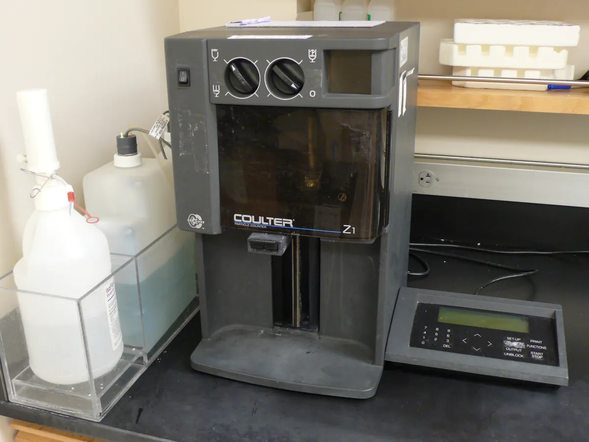
Most automated counters are image-based. A small volume of sample mixed with trypan blue (bright-field) or with fluorescent dyes such as acridine orange and propidium iodide (AO/PI) is loaded into a chamber of known depth. A camera images a defined volume, and software finds each cell, measures its size and brightness, and classifies it as live or dead. Other types use electrical impedance (Coulter principle) to count and size particles without imaging.
Because the result depends on the counting volume, focus and the software's classification settings, counters are checked with certified reference beads and compared with manual counts. ISO 20391 gives a framework for evaluating cell counting methods.
Used for
- Routine cell counts and viability during passaging
- Seeding density for bioreactors and assays
- In-process monitoring of fed-batch and perfusion cultures
- Cell bank and cell therapy product release testing (with a validated method)
- Cell size distribution and aggregation checks
How it works
- 01
Mix
Mix the cell suspension well so the sample is representative.
- 02
Stain
Mix a set volume of sample with stain (for example 1:1 with 0.4% trypan blue).
- 03
Load
Load the mixture into the counting chamber without bubbles.
- 04
Focus and image
The instrument focuses automatically and images one or more fields of known volume.
- 05
Classify
Software identifies objects, applies size and brightness gates, and classifies them live or dead.
- 06
Report
Live cells/mL, total cells/mL, viability %, mean size and aggregation are reported, corrected for dilution.
Advantages
- Faster than manual counting: typically under a minute per sample
- Less operator-to-operator variation
- Counts a larger volume than a typical manual count, improving precision
- Stores images and data for review and audit trail
- Fluorescent AO/PI methods work better than trypan blue for samples with debris or red cells
Limitations
- Cell clumps can be counted as one object or mis-sized
- Debris and dead-cell fragments can be miscounted as cells
- Classification depends on software gates that must be set and locked for each cell type
- Trypan blue is toxic to cells over time, so stained samples must be counted promptly
- Disposable slides add running cost
1 calibration procedure
Methods that use Cell counter
Keep every Cell counter in control: usage logs, calibration and maintenance schedules, and automatic blocking of overdue instruments.