Viable Cell Count and Viability by Trypan Blue Exclusion
A well-mixed cell suspension is mixed 1:1 with 0.4% trypan blue. Unstained (live) and blue (dead) cells are counted in a haemocytometer or an automated counter. Viable cells/mL and per cent viability are calculated using the chamber volume factor and the dilution factor.
- Biotech & biologics
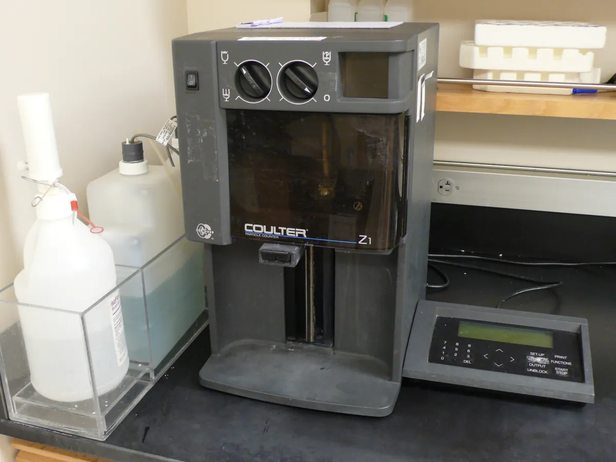
- Trypan blue / automated counter
- Cell counter
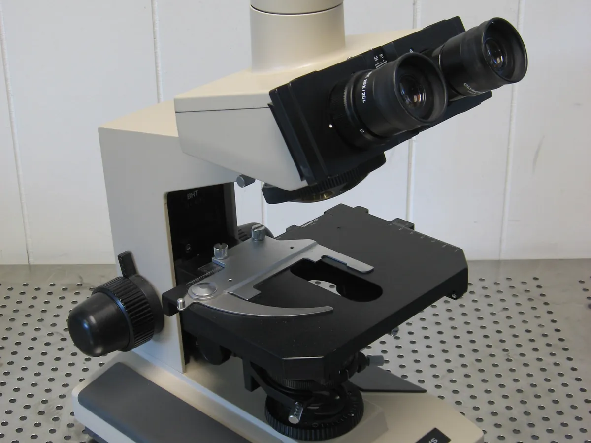
- Optical microscope
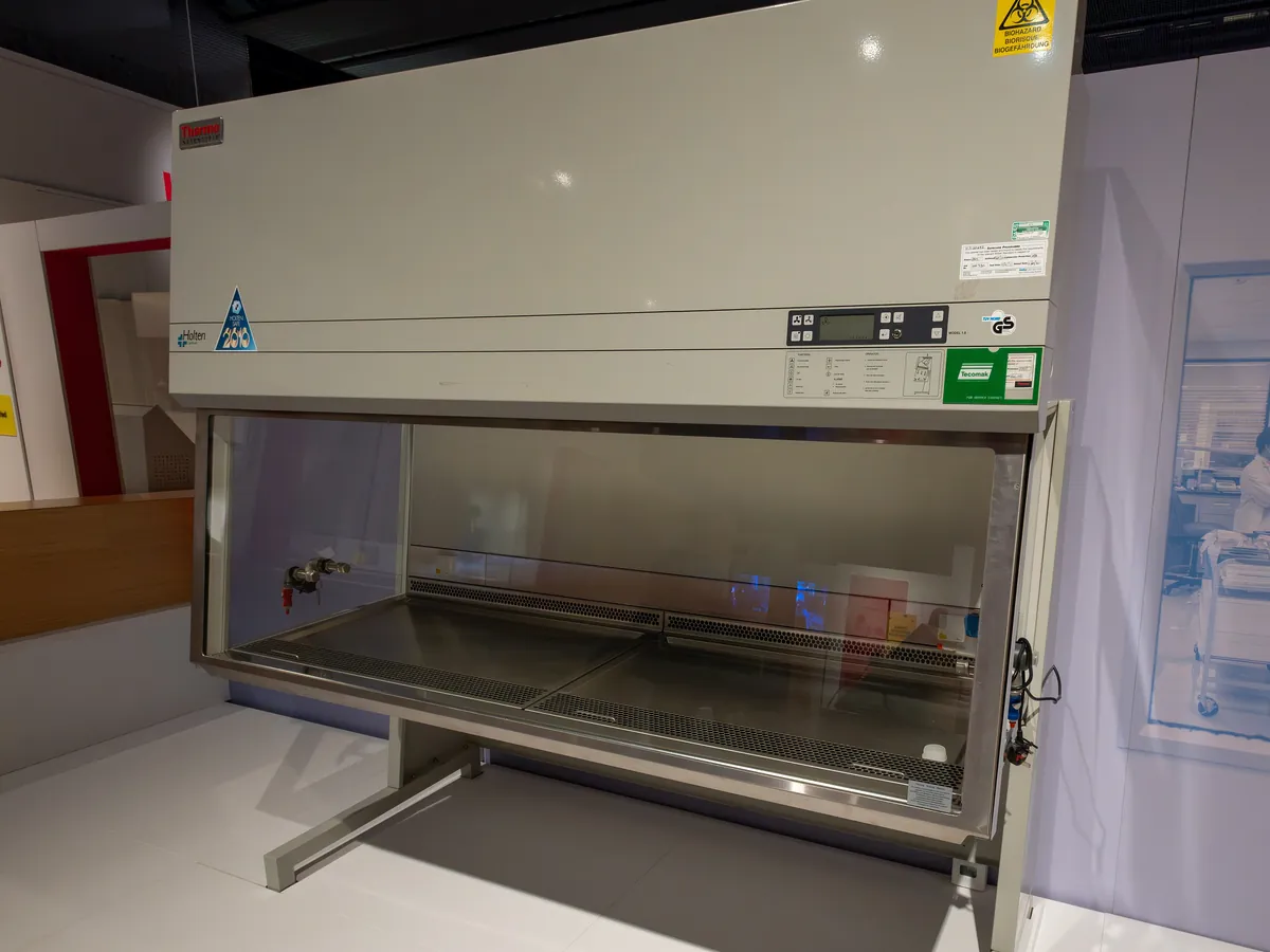
- BSC
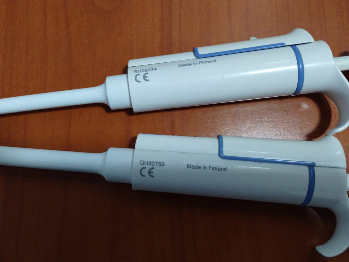
- Micropipette
Reference: Typical method based on Ph. Eur. 2.7.29 (dye exclusion) and the principles of ISO 20391-1. Use the validated procedure for your cell type and counting system.
Why this method works
Live cells have intact membranes and exclude trypan blue, so they appear bright and clear. Dead cells with damaged membranes take up the dye and appear blue. In a Neubauer haemocytometer, each large corner square is 1 mm × 1 mm and the chamber depth is 0.1 mm, so each square holds 0.1 mm³ = 10⁻⁴ mL. The average count per large square is therefore multiplied by 10⁴ to give cells per mL, and then by the dilution factor from mixing with stain. Viability is the live count as a percentage of the total (live + dead) count. Automated counters apply the same calculation to a known imaged volume.
Instruments used
Reagents & standards
| Reagent | Grade / specification |
|---|---|
| Trypan blue | 0.4% (w/v) in buffered saline, filtered to remove crystals |
| Diluent (if needed) | Phosphate-buffered saline or culture medium to bring cells into the countable range |
Preparing the sample
- 01
Mix culture
Swirl or gently pipette the culture so cells are evenly suspended.
- 02
Sample
Take a representative sample aseptically in the biosafety cabinet.
- 03
Stain
Mix equal volumes of cell suspension and 0.4% trypan blue (DF = 2).
- 04
Load
Load the haemocytometer or counter slide immediately.
- 05
Count
Count live and dead cells in the defined squares within a few minutes.
- 06
Calculate
Calculate viable cells/mL, total cells/mL and viability.
Procedure
- 1Clean the haemocytometer and cover slip with 70% ethanol, dry, and place the cover slip over the counting area.
- 2Gently mix the cell suspension by pipetting or inversion and take a sample at once, before cells settle.
- 3If the culture is dense, dilute it with PBS or medium first and record the extra dilution.
- 4Mix 20 µL of cell suspension with 20 µL of 0.4% trypan blue (dilution factor 2). Mix gently by pipetting.
- 5Load about 10 µL into each side of the haemocytometer by capillary action. Do not overfill or leave bubbles.
- 6Under the microscope (10× objective), count live (clear) and dead (blue) cells in the four large corner squares on one side. Count cells touching the top and left lines but not the bottom and right lines.
- 7Aim for about 100–500 cells in total for good precision; adjust the dilution if counts are far outside this range.
- 8Count the second side or repeat the count as required by the method, and complete within about 3–5 minutes of staining.
- 9For an automated counter, load the slide with the same mix and use the validated protocol with dilution factor 2.
Notes for analysts
- Trypan blue slowly enters live cells too, so long exposure gives falsely low viability. Count promptly.
- Serum proteins bind trypan blue and can raise background; for heavily serum-containing samples, wash cells or use the method's adjustment.
- Clumped cells make counting unreliable. Mix gently to separate them or use an enzymatic or declumping step if validated.
- Viability by dye exclusion only shows membrane integrity; it does not show whether cells can still divide or function.
Method parameters (typical)
| Stain | 0.4% trypan blue, mixed 1:1 with cells (DF = 2) |
|---|---|
| Chamber | Improved Neubauer haemocytometer, depth 0.1 mm; or automated counter slide |
| Squares counted | 4 large corner squares (each 1 mm², volume 10⁻⁴ mL) |
| Target count | About 25–125 cells per large square (about 100–500 in total) |
| Time from staining to count | Within about 3–5 minutes |
System suitability & sequence
| Parameter | Limit |
|---|---|
| Counter verification | Automated counter within its bead check date |
| Replicate agreement | Duplicate counts typically within about 10–15% of each other |
| Count range | Cells per square within the target range; otherwise re-dilute |
Typical sequence
- 1Clean and prepare chamber
- 2Mix culture and sample
- 3Stain 1:1 with trypan blue
- 4Load chamber
- 5Count live and dead cells
- 6Repeat count
- 7Calculate and report
Calculation with a worked example
Where
- live
- Unstained cells counted in all squares
- dead
- Blue cells counted in all squares
- squares
- Number of large squares counted (here 4)
- DF
- Dilution factor from mixing with trypan blue (and any pre-dilution); here 2
- 10⁴
- Haemocytometer factor: one large square holds 10⁻⁴ mL
Given
- Live cells per square
- 112, 98, 105, 109 (total 424)
- Dead cells per square
- 6, 8, 5, 7 (total 26)
- Dilution
- 1:1 with trypan blue (DF = 2)
Working
- 1Mean live cells per square = 424 ÷ 4 = 106
- 2
Viable cells/mL = 106 × 2 × 10⁴ = 2.12 × 10⁶ cells/mL
- 3Total cells/mL = (450 ÷ 4) × 2 × 10⁴ = 112.5 × 2 × 10⁴ = 2.25 × 10⁶ cells/mL
- 4Viability = 424 ÷ (424 + 26) × 100 = 424 ÷ 450 × 100 = 94.2%
Viable cell density = 2.12 × 10⁶ cells/mL; viability = 94.2%
What result passes
- Compare with the in-process or release criteria for the culture (for example many cell banks and seed cultures require viability of at least about 80–90%; use the limits in your specification).
- Duplicate counts within the agreement limit in the method.
Validation parameters (ICH Q2(R2))
| Parameter | Typical requirement |
|---|---|
| Accuracy | Comparison of automated and manual counts; bead standards for the counter |
| Precision | Repeatability and intermediate precision across operators and days (CV typically ≤ 10–15%) |
| Linearity | Dilution series of cells across the working range (ISO 20391-2 approach) |
| Range | Lowest and highest densities that meet precision and linearity criteria |
| Robustness | Time in stain, mixing and stain lot |
Run this method in LimsPulse: specifications and STPs under version control, results calculated and checked against limits, and a signed Certificate of Analysis.