Residual host cell DNA by quantitative PCR (qPCR)
Measures the very small amount of DNA from the host cells left in the purified protein. DNA is extracted from the sample, a sequence that occurs many times in the host genome is amplified by qPCR, and the amount is read from a standard curve of host-cell genomic DNA. The result is reported per mg of product and per dose; a commonly applied limit is 10 ng per dose (typical, from WHO guidance).
- Biotech & biologics
- DNA extraction followed by real-time quantitative PCR against a genomic DNA standard curve
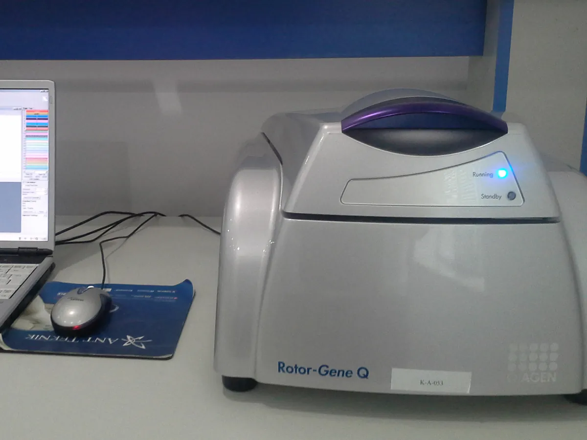
- qPCR
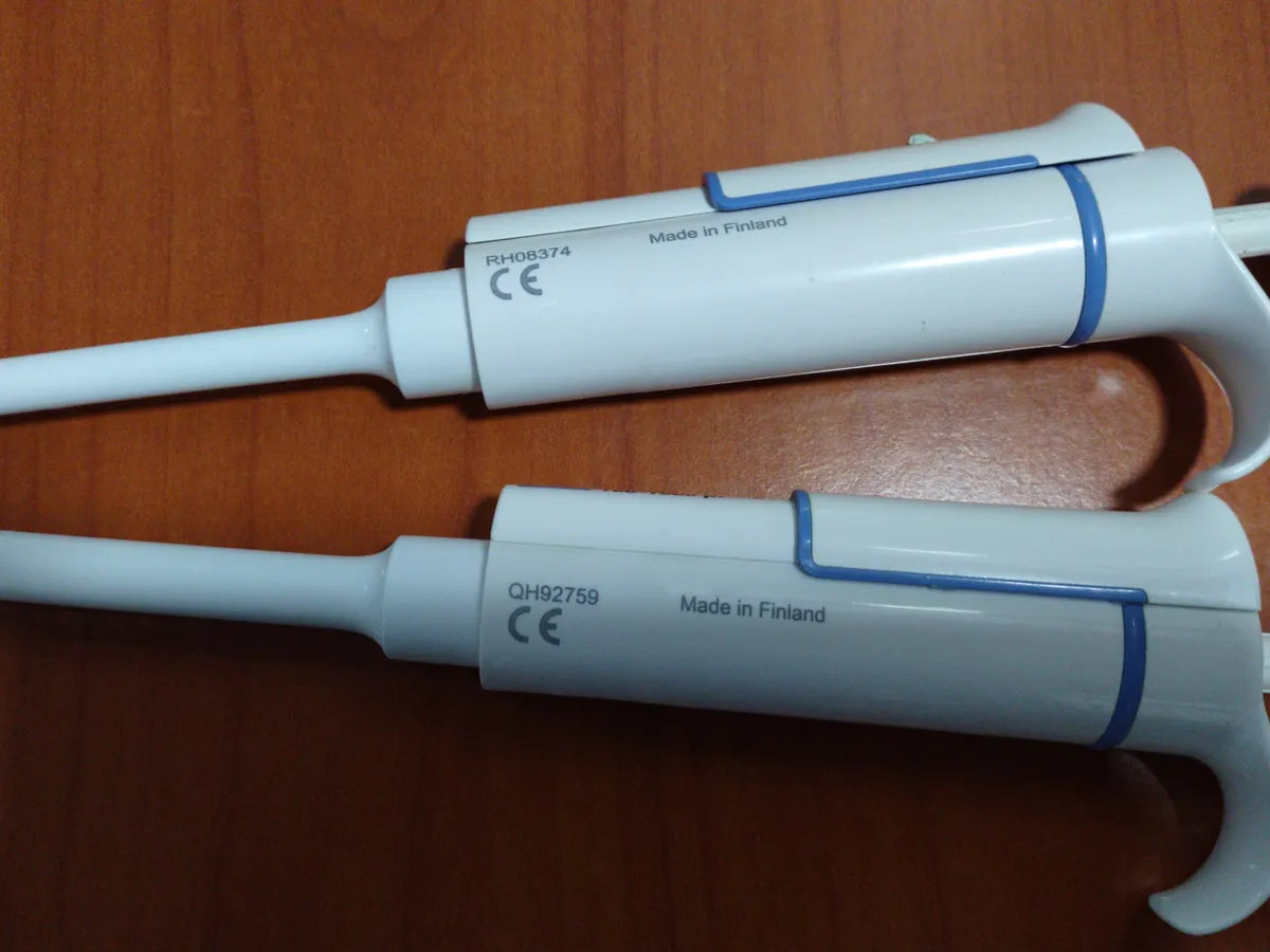
- Micropipette
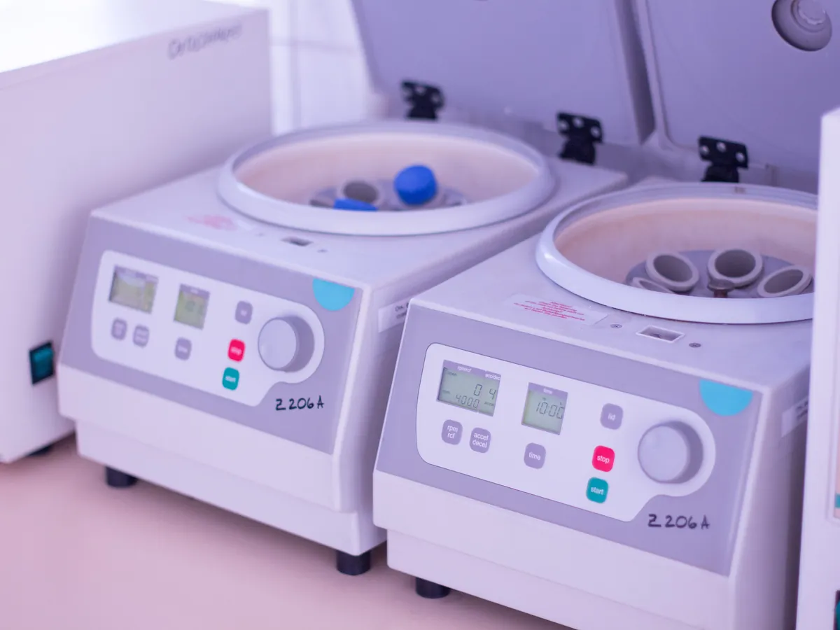
- Centrifuge
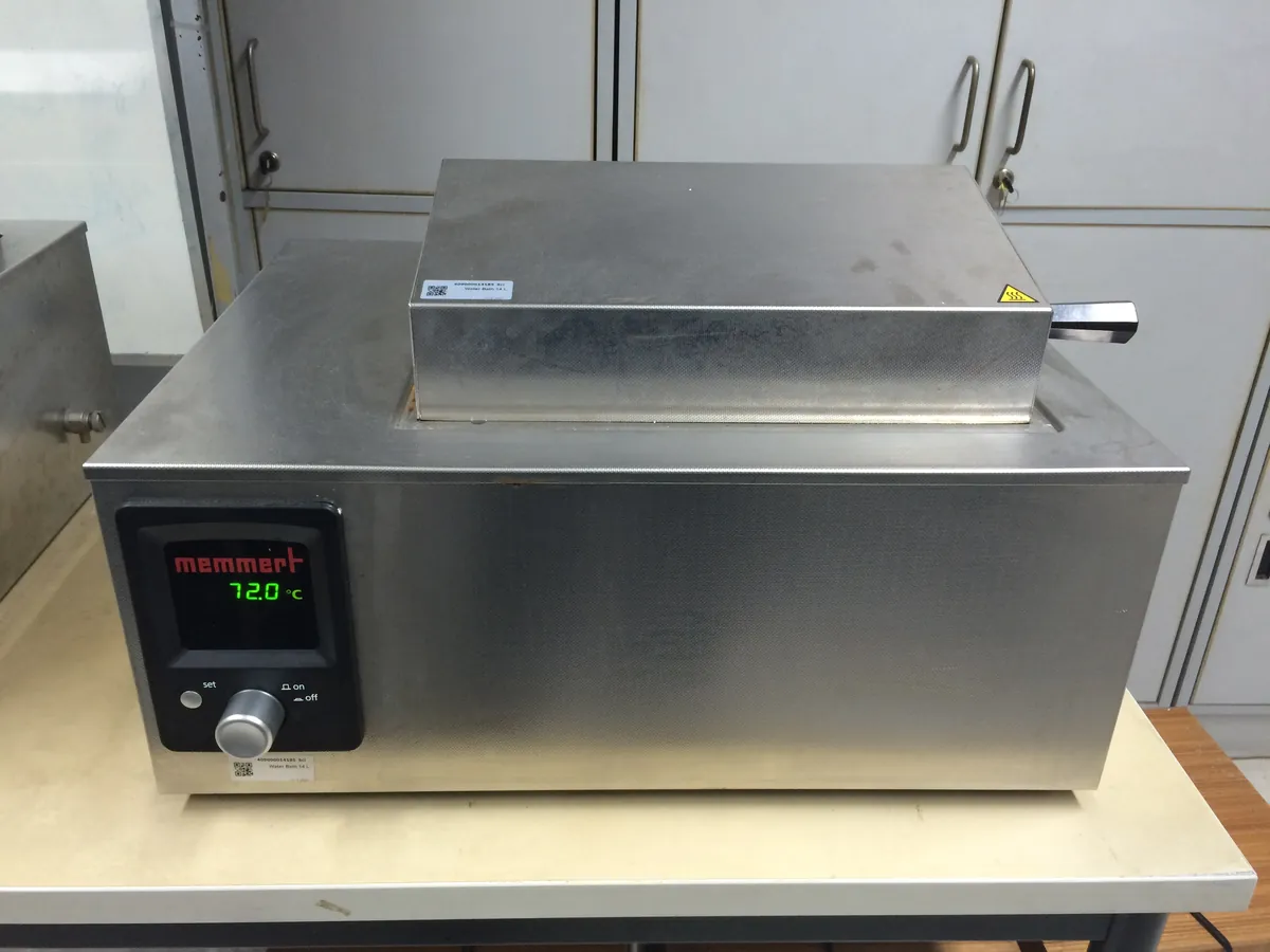
- Water bath
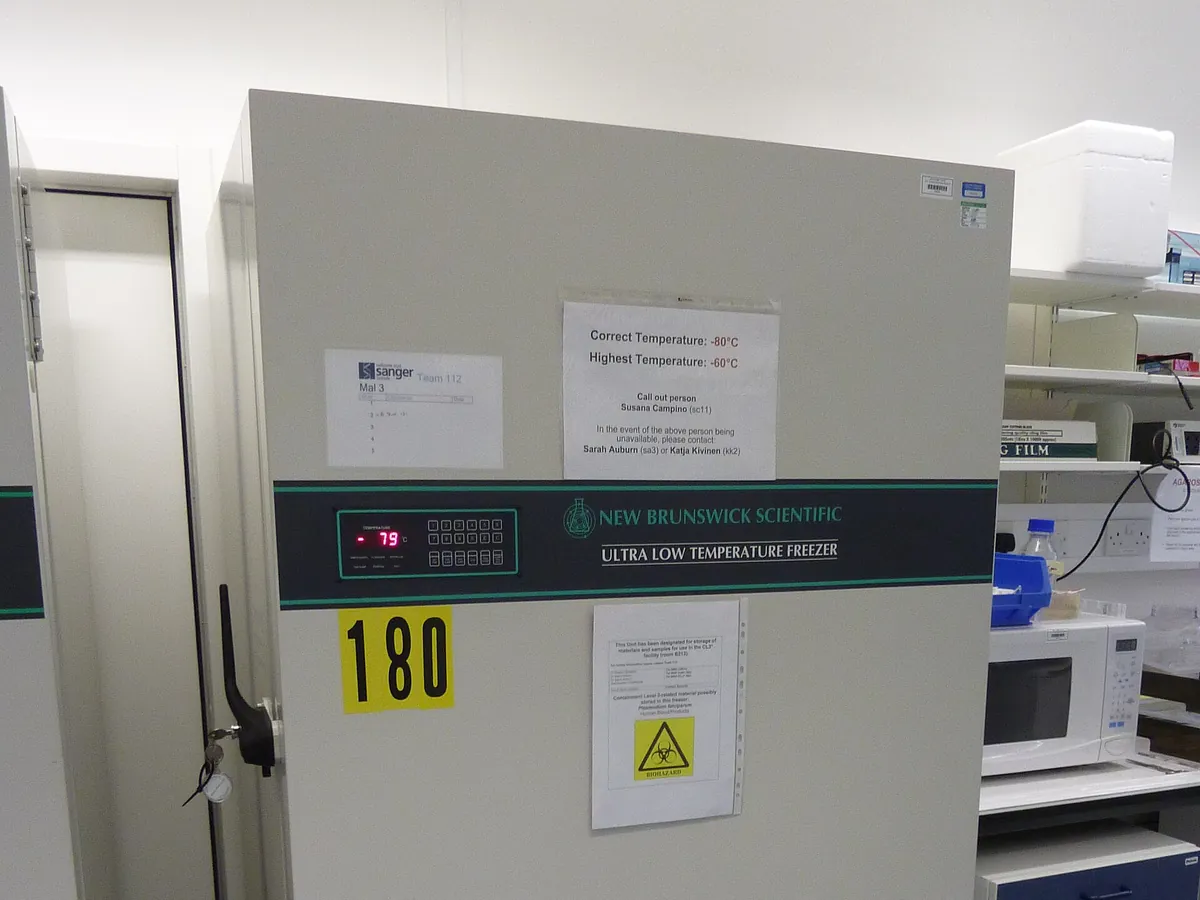
- Fridge / freezer
Reference: Typical method following the approach of USP <509>, USP <1130> and WHO guidance on cell substrates. Use the kit or in-house assay and limits in your approved procedure.
Why this method works
Real-time PCR copies a short target DNA sequence in repeated heating and cooling cycles, roughly doubling it each cycle. A fluorescent probe or dye reports the amount of product after every cycle. The cycle at which fluorescence crosses a set threshold is the quantification cycle (Cq, also called Ct). The more target DNA at the start, the earlier the curve crosses the threshold: a 10-fold increase in starting DNA lowers Cq by about 3.32 cycles when efficiency is 100%. Plotting Cq against log10 of the amount for a dilution series of host-cell genomic DNA gives a straight-line standard curve, and unknowns are read from it. Assays target highly repeated sequences in the host genome (for example repetitive elements) so that femtogram-to-picogram amounts can be detected. Because protein, salts and detergents in the sample can inhibit PCR, the DNA is first extracted (for example proteinase K digestion followed by magnetic-bead or column purification), and a spiked extraction control measures how much DNA is recovered.
Instruments used
Reagents & standards
| Reagent | Grade / specification |
|---|---|
| Host-cell genomic DNA standard | Purified genomic DNA of the production cell line, concentration assigned (e.g. by A260 or fluorescent dye) |
| Primers and probe | Specific for a repetitive host-genome target (kit or in-house), probe with fluorophore and quencher |
| qPCR master mix | DNA polymerase, dNTPs, buffer and Mg²⁺; may include uracil-DNA glycosylase to limit carry-over |
| Extraction reagents | Proteinase K, lysis buffer, magnetic beads or spin columns, wash and elution buffers |
| Carrier / diluent | DNA-free water or dilution buffer, sometimes with carrier nucleic acid |
| Controls | No-template control (NTC), extraction blank, spiked extraction control, internal positive control if used |
Preparing the sample
- 01
Separate areas
Set up reagents in a clean area, away from DNA standards and products.
- 02
Spike
Spike one sample aliquot with a known amount of genomic DNA.
- 03
Digest
Digest protein with proteinase K at the validated temperature.
- 04
Extract
Purify DNA by magnetic beads or columns; elute in a small volume.
- 05
Set up qPCR
Add eluates, standards and controls to the master mix in the plate.
- 06
Run & analyse
Run the cycling program and read Cq against the standard curve.
Procedure
- 1Work in a PCR-clean area with dedicated pipettes and filter tips. Keep genomic DNA standards and amplified products away from reagent preparation to avoid contamination.
- 2Take a fixed volume of sample (e.g. 200 µL). Prepare a second aliquot spiked with a known amount of host genomic DNA as the extraction recovery control, and an extraction blank of DNA-free water.
- 3Add lysis buffer and proteinase K and incubate in a water bath (or heat block) at the validated temperature (typically about 50–60 °C) to digest the protein.
- 4Purify the DNA by magnetic-bead or column extraction, wash, and elute into a small volume (e.g. 50 µL), which concentrates it.
- 5In the qPCR plate, add master mix with primers and probe, then the genomic DNA standard series, sample eluates, spiked eluate, extraction blank and NTC, usually in triplicate.
- 6Seal, spin briefly and run on the qPCR system using the validated cycling program (an initial activation step then about 40 cycles of denaturation and annealing/extension).
Standard preparation
- 1Prepare a 10-fold dilution series of host-cell genomic DNA in the validated diluent, typically 5 or more levels (for example 300, 30, 3, 0.3 and 0.03 pg per reaction).
- 2Prepare fresh dilutions or use validated frozen aliquots; avoid repeated freeze–thaw.
- 3Run the standards in triplicate on every plate.
Notes for analysts
- Because only a repetitive target is amplified, the result is 'DNA equivalent' to the genomic standard. Very short DNA fragments (below the amplicon length) may not be counted; some regulators also ask about fragment size.
- Contamination is the main risk: one stray microlitre of concentrated genomic DNA can ruin weeks of work. Keep pre- and post-PCR areas separate and never open used PCR plates in the setup area.
- Older methods (hybridisation, threshold total-DNA assay) exist; qPCR is now the most common because it is sensitive and specific to the host species.
Method parameters (typical)
| Target | Repetitive sequence in the host cell genome (kit- or method-specific) |
|---|---|
| Chemistry | Hydrolysis (TaqMan-type) probe or validated intercalating dye |
| Reaction volume | e.g. 25–30 µL including 5–10 µL eluate (as validated) |
| Cycling | Polymerase activation, then about 40 cycles of denaturation (~95 °C) and annealing/extension (~60 °C) |
| Standard curve | At least 5 levels covering the reporting range, triplicate |
| Reporting | pg DNA per mg product and per dose |
System suitability & sequence
| Parameter | Limit |
|---|---|
| Standard curve slope | Typically about −3.1 to −3.6 (amplification efficiency about 90–110%) |
| Correlation (R²) | Typically ≥ 0.98 (or as stated in the procedure) |
| No-template control | No amplification, or Cq later than the lowest standard by the stated margin |
| Extraction blank | Below the quantitation limit |
| Spike recovery | Within the validated range, commonly 50–150% for residual DNA assays |
| Replicate precision | Cq standard deviation (or %CV of quantity) within the stated limit |
Typical sequence
- 1Standard curve (5 levels) in triplicate
- 2No-template control in triplicate
- 3Extraction blank in triplicate
- 4Sample eluate in triplicate
- 5Spiked sample eluate in triplicate
Calculation with a worked example
Where
- Cq
- Mean quantification cycle of the sample
- m, b
- Slope and intercept of the standard curve (Cq against log10 pg)
- Q
- DNA per reaction (pg) read from the curve
- V(sample per reaction)
- Original sample volume represented in one reaction (mL)
- V(dose)
- Volume of drug substance making up one dose (mL)
Given
- Standard curve
- m = −3.32, b = 27.5 (Cq of 1 pg)
- Sample mean Cq
- 28.4
- Extraction
- 200 µL sample eluted in 50 µL; 5 µL eluate per reaction = 20 µL (0.020 mL) of sample
- Product
- 5 mg/mL; dose 100 mg = 20 mL
Working
- 1Efficiency = (10^(1/3.32) − 1) × 100 = (2.0008 − 1) × 100 = 100.1%
- 2log10 Q = (28.4 − 27.5) / (−3.32) = −0.2711, so Q = 10^−0.2711 = 0.536 pg per reaction
- 3
DNA in sample = 0.536 pg / 0.020 mL = 26.8 pg/mL
- 4
Per mg = 26.8 / 5 = 5.36 pg/mg
- 5
Per dose = 26.8 pg/mL × 20 mL = 536 pg = 0.54 ng/dose
Residual host cell DNA = 5.4 pg/mg, about 0.54 ng per 100 mg dose — well below a typical 10 ng/dose limit
What result passes
- Residual DNA not more than the product specification limit, usually expressed per dose. A commonly applied limit for products made in continuous cell lines is 10 ng per dose (typical, from WHO guidance); some products justify other limits based on risk and DNA fragment size.
- All run controls (standard curve, NTC, extraction blank, spike recovery) meet their criteria; otherwise the run is invalid.
- Results below the quantitation limit reported as less than the LOQ, expressed per mg and per dose.
Validation parameters (ICH Q2(R2))
| Parameter | Typical requirement |
|---|---|
| Specificity | Primers amplify the host target only; no signal from product, other cell lines or reagents |
| Accuracy | Spike recovery of genomic DNA through extraction in the product matrix |
| Linearity and range | Standard curve linear over at least 4–5 logs |
| Precision | Repeatability and intermediate precision of quantity, including extraction |
| LOD / LOQ | Lowest DNA amount detected / quantified reliably, typically femtogram to low-picogram per reaction |
| Robustness | Annealing temperature, master mix lot, extraction operator and incubation time |
Run this method in LimsPulse: specifications and STPs under version control, results calculated and checked against limits, and a signed Certificate of Analysis.