Optical (Light) Microscope
Magnifies particles so their size, shape and crystal habit can be seen and measured with a calibrated eyepiece graticule or camera. Used for particle size, morphology and identification checks.
- Pharmaceutical
- Microbiology
- Materials
- Food
- Forensics
- Education
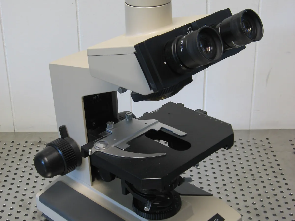
What is Optical microscope?
A compound optical microscope uses two lens systems, the objective and the eyepiece, to give magnifications from about 40× to 1000×. With a camera and image software it becomes a simple particle imaging system.
In pharmaceutical QC it is used to look at particle shape and crystal habit, to check for agglomerates, to estimate particle size with a calibrated graticule, and to confirm results from laser diffraction. With crossed polarisers it shows whether particles are crystalline (they glow, or show birefringence) or amorphous.
Microscopy counts individual particles, so it gives a number-based size distribution, unlike the volume-based results from laser diffraction. It is also used in microbiology (Gram stain), for suspension checks and for identifying foreign particles found in products.
Used for
- Particle size and shape of APIs and excipients
- Crystallinity check with polarised light (birefringence)
- Checking suspensions for crystal growth on stability
- Identification of foreign particles in products
- Supporting laser diffraction results (agglomerates, needles)
- Microscopic particle count method for injections (as an alternative to light obscuration)
How it works
- 01
Illumination
A lamp and condenser send an even cone of light through the slide.
- 02
Sample
Particles are dispersed in a suitable mounting fluid on a glass slide with a cover slip.
- 03
Objective
Forms a magnified real image of the particles (e.g. 4×, 10×, 40×, 100×).
- 04
Eyepiece / camera
Magnifies the image further (usually 10×) or projects it onto a camera.
- 05
Measurement
Particles are sized against a graticule calibrated with a stage micrometer.
Advantages
- Direct visual information on shape and habit
- Low cost and easy to use
- Small sample needed
- Polarised light distinguishes crystalline and amorphous material
Limitations
- Only a small number of particles is examined, so sampling errors are large
- Manual sizing is slow and depends on the analyst
- Practical lower limit about 1 µm
- Gives a 2D projection of 3D particles
Methods that use Optical microscope
Keep every Optical microscope in control: usage logs, calibration and maintenance schedules, and automatic blocking of overdue instruments.