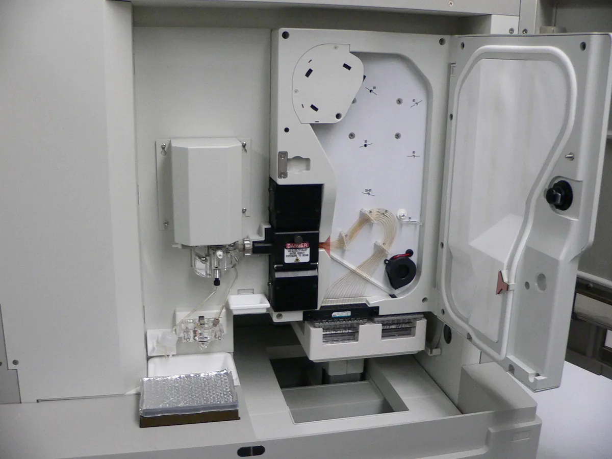
Capillary electrophoresis system
Separates charged molecules in a thin fused-silica capillary under a high voltage (typically up to 30 kV). Used in biologics QC for size purity (CE-SDS), charge variants (cIEF, CZE) and glycan analysis.
- Biotechnology
- Biopharmaceutical
- Pharmaceutical
- Genomics
- Forensic science
- Academic research
How CE works
When a voltage is applied, ions move with an electrophoretic mobility that depends on charge and size. In an uncoated fused-silica capillary, the negatively charged wall also drives a bulk flow of liquid called electroosmotic flow (EOF). The observed migration is the sum of both. In CE-SDS, a sieving polymer and SDS make separation depend on size; in cIEF, proteins migrate in a pH gradient until they reach their isoelectric point and are then mobilised past the detector.
The path of a sample
- 01
Condition
The capillary is rinsed with conditioning solutions and filled with background electrolyte or sieving gel.
- 02
Inject
A small plug of sample is introduced by pressure or by voltage (electrokinetic injection).
- 03
Apply voltage
High voltage is applied across the capillary ends dipping in buffer vials.
- 04
Separate
Molecules migrate at different speeds towards the detector window.
- 05
Detect
UV (often 214 or 220 nm, 280 nm for cIEF) or laser-induced fluorescence detects analytes through a clear window in the capillary.
- 06
Integrate
Software integrates peaks and calculates corrected peak areas and relative per cent.
Main parts and what they do
Capillary and cartridge
Fused-silica capillary (bare or coated) in a cartridge that is thermostatted by liquid or air cooling.
High-voltage power supply
Applies up to about ±30 kV with precise control of voltage or current.
Sample and buffer trays
Hold vials for samples, buffers, rinses and waste; often temperature controlled.
Injection system
Applies pressure or voltage to introduce a defined sample plug.
Detector
UV/PDA or laser-induced fluorescence (LIF) at a window in the capillary.
Software
Controls methods, collects electropherograms, integrates peaks and keeps an audit trail.
Terms every analyst should know
Electrophoretic mobility
Speed per unit field strength. Proportional to charge and inversely related to size (hydrodynamic radius).
Electroosmotic flow (EOF)
Bulk flow caused by the charged capillary wall. Coated capillaries suppress EOF in CE-SDS and cIEF.
Joule heating
Heat generated by current. The thermostatted cartridge removes it; poor cooling broadens peaks and shifts migration times.
Ohm's law plot
Current plotted against voltage. It is straight while heat is removed effectively; a curve upwards shows excess Joule heating.
Corrected peak area
Area divided by migration time. Slower species spend longer in the detector window, so correction is needed for accurate relative per cent.
Isoelectric point (pI)
The pH at which a protein has no net charge. cIEF separates charge variants by pI using pI markers for calibration.
Keep every CE in control: usage logs, calibration and maintenance schedules, and automatic blocking of overdue instruments.