Automated cell counter
Counts cells and measures their viability automatically, usually by imaging a stained sample in a disposable or reusable counting chamber. It replaces manual haemocytometer counting for routine cell culture and bioprocess work.
- Biotechnology
- Biopharmaceutical
- Cell and gene therapy
- Academic research
- Vaccine manufacture
- Regenerative medicine
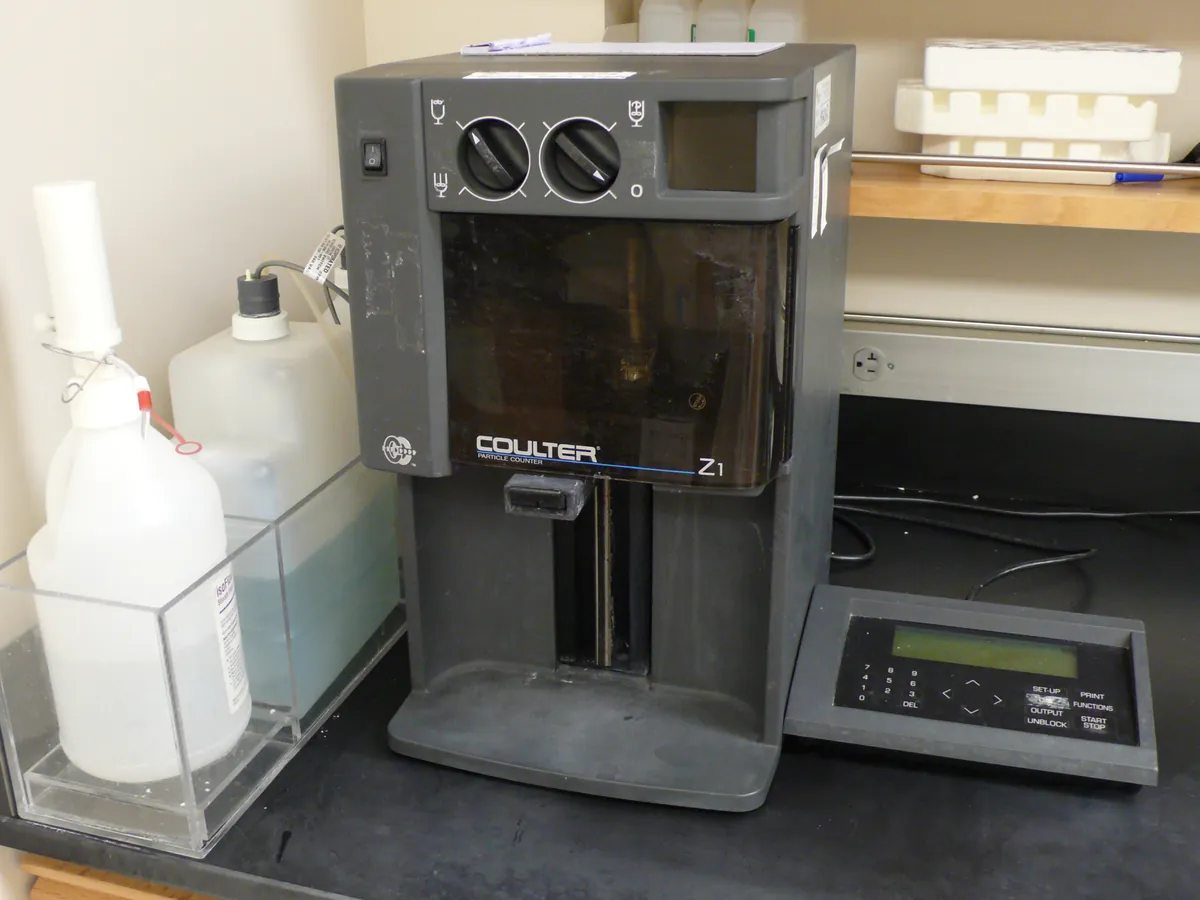
How Cell counter works
Trypan blue is excluded by cells with intact membranes, so live cells look bright and dead cells look blue. In AO/PI mode, acridine orange enters all nucleated cells (green) and propidium iodide enters only cells with damaged membranes (red). The counter images a known volume, counts live and dead objects and calculates concentration = count ÷ volume imaged × dilution factor, and viability = live ÷ (live + dead) × 100.
The path of a sample
- 01
Mix
Mix the cell suspension well so the sample is representative.
- 02
Stain
Mix a set volume of sample with stain (for example 1:1 with 0.4% trypan blue).
- 03
Load
Load the mixture into the counting chamber without bubbles.
- 04
Focus and image
The instrument focuses automatically and images one or more fields of known volume.
- 05
Classify
Software identifies objects, applies size and brightness gates, and classifies them live or dead.
- 06
Report
Live cells/mL, total cells/mL, viability %, mean size and aggregation are reported, corrected for dilution.
Main parts and what they do
Counting chamber or slide
Holds a fixed volume of sample at a known depth; disposable or reusable.
Illumination
Bright-field LED for trypan blue; LED excitation with filters for fluorescence modes.
Optics and camera
Objective and image sensor with autofocus that images the chamber.
Image analysis software
Finds and classifies cells using size, brightness and roundness settings; stores images.
Protocol library
Locked settings per cell type, with user access control in GMP versions.
Terms every analyst should know
Dye exclusion
Live cells with intact membranes keep dyes such as trypan blue or propidium iodide out. Dead cells take them up.
Dilution factor
Mixing cells 1:1 with stain gives a dilution factor of 2. The software must use the same factor as the actual mix.
Size gate
Minimum and maximum diameter limits so debris and clumps are excluded. They are set for each cell type and locked in the protocol.
Counting statistics
The relative error of a count is roughly 1 ÷ √N. Counting 400 cells gives about 5% relative error; counting 100 gives about 10%.
Reference beads
Polystyrene beads of certified concentration and size, used to check counting accuracy, precision and size measurement.
Keep every Cell counter in control: usage logs, calibration and maintenance schedules, and automatic blocking of overdue instruments.