Bioanalytical
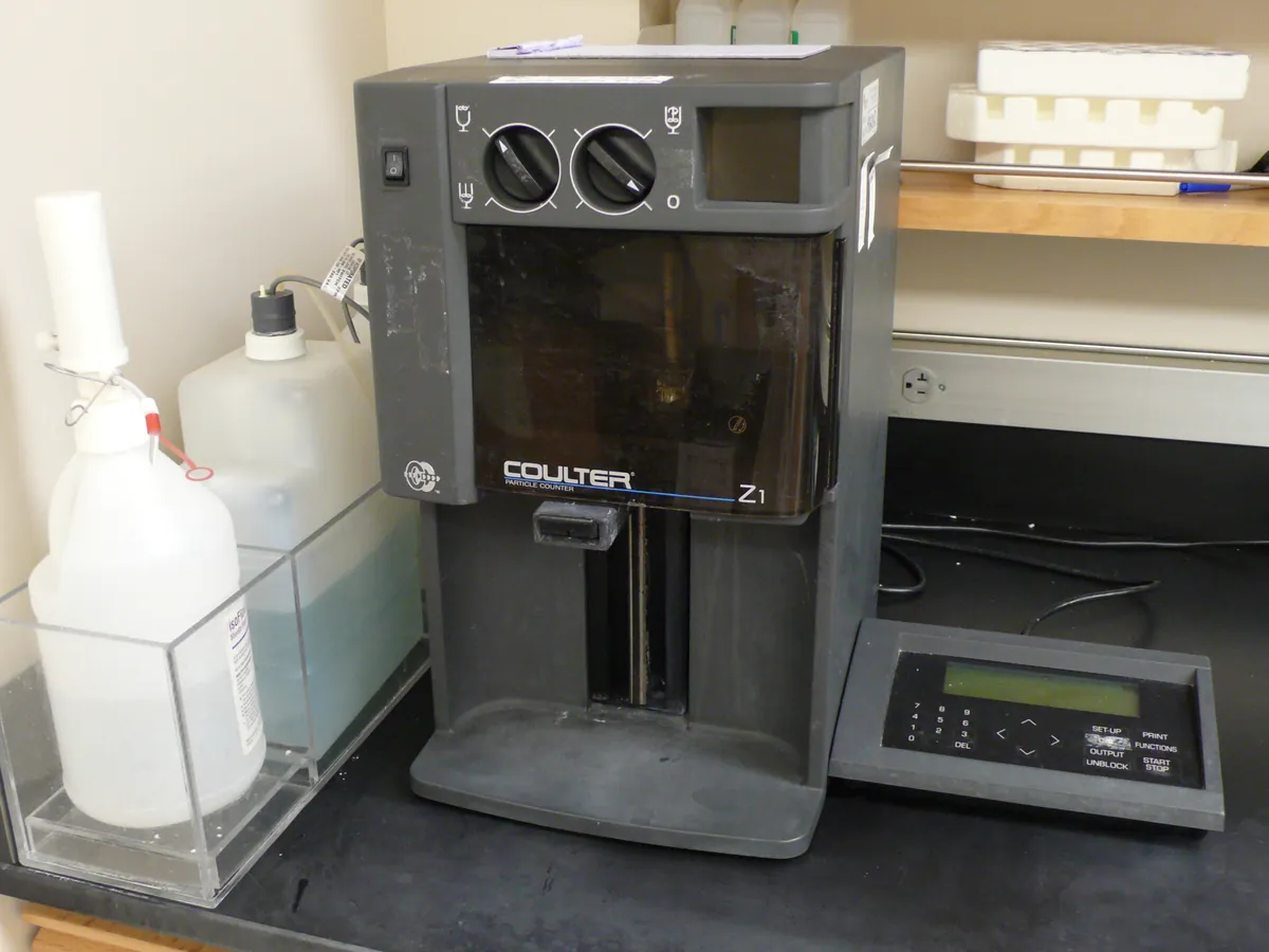
Automated cell counter
Counts cells and measures their viability automatically, usually by imaging a stained sample in a disposable or reusable counting chamber. It replaces manual haemocytometer counting for routine cell culture and bioprocess work.
- Biotechnology
- Biopharmaceutical
- Cell and gene therapy
- Academic research
- Vaccine manufacture
- Regenerative medicine
TROUBLESHOOTING
Common problems, causes and fixes
Viability lower than expected
Likely causes
- • Cells left too long in trypan blue
- • Rough handling or cell stress
- • Debris classified as dead cells
What to do
- • Count within the set time after staining
- • Mix gently; check culture conditions
- • Review size gates and the image overlay
Counts vary widely between replicates
Likely causes
- • Cells settled before sampling
- • Clumps
- • Bubbles or poorly filled chamber
What to do
- • Mix immediately before sampling
- • Gently pipette to break clumps or use a declumping setting
- • Reload the chamber carefully
Counts differ from manual haemocytometer
Likely causes
- • Different size gates or classification
- • Wrong dilution factor entered
- • Out-of-range concentration
What to do
- • Align gates with the manual definition of a cell
- • Check the dilution factor in the protocol
- • Dilute or concentrate into the validated range
Image out of focus or fails to focus
Likely causes
- • Dirty slide or optics
- • Slide not seated properly
What to do
- • Clean optics and use a new slide
- • Reinsert the slide fully
Bead count outside certificate range
Likely causes
- • Beads not resuspended
- • Expired beads
- • Instrument fault
What to do
- • Vortex beads well before use
- • Use in-date beads
- • Contact service and do not use for reportable counts
In LimsPulse · Calibration Management
Keep every Cell counter in control: usage logs, calibration and maintenance schedules, and automatic blocking of overdue instruments.