Thin-Layer Chromatography (TLC)
Separates compounds as a solvent climbs up a thin layer of adsorbent on a glass, aluminium or plastic plate. Simple and cheap; used in pharma QC mainly for identification and limit tests for related substances.
- Pharmaceutical
- Herbal & natural products
- Chemicals
- Forensic
- Education
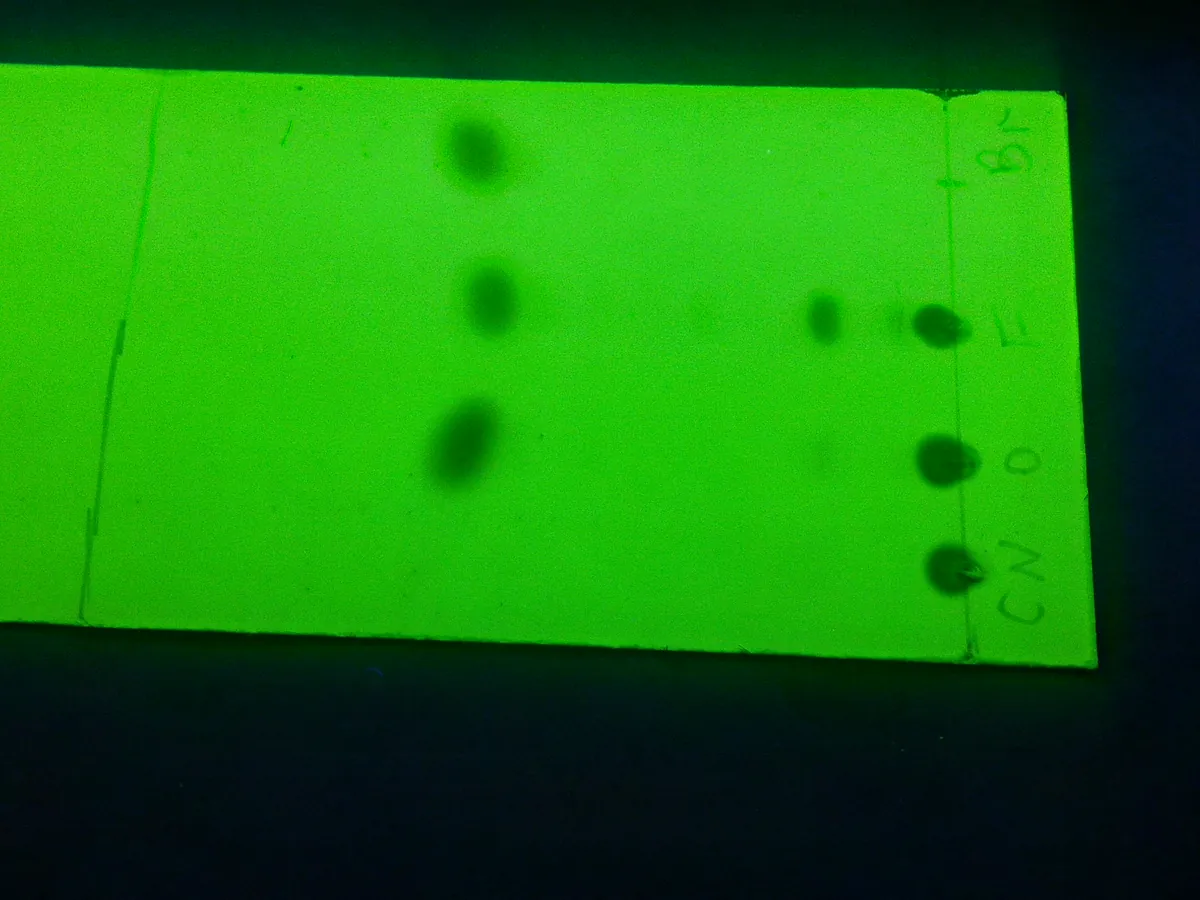
What is TLC?
A TLC plate is coated with a thin, even layer of adsorbent, most often silica gel. Small spots of sample and reference solutions are applied near the bottom edge. The plate is placed in a closed tank containing a shallow layer of solvent (the mobile phase).
The solvent rises up the plate by capillary action. Compounds move with it at different speeds depending on how strongly they stick to the silica compared with how well they dissolve in the solvent. When the solvent front is near the top, the plate is removed, dried and the spots are made visible (visualisation).
Each spot is described by its Rf value: the distance moved by the spot divided by the distance moved by the solvent front. Identification compares the sample spot's position, colour and size with a reference standard run on the same plate. Limit tests compare the intensity of impurity spots with a diluted reference solution.
Used for
- Identification of APIs and excipients against a reference standard
- Limit tests for related substances in older pharmacopoeial monographs
- Identification of herbal materials (fingerprinting)
- Monitoring reaction progress in synthesis
- Quick screening of cleaning samples or counterfeit products
How it works
- 01
Prepare chamber
Add mobile phase to a depth below the application line, line the tank with filter paper and close it to saturate (often 30 minutes or as the method states).
- 02
Apply spots
Apply small, equal volumes of test and reference solutions on a pencil line about 1.5–2 cm from the bottom edge.
- 03
Develop
Place the plate in the tank and let the solvent rise to a marked distance (e.g. 10–15 cm or three-quarters of the plate).
- 04
Dry
Remove the plate, mark the solvent front immediately and dry in air or a stream of warm air.
- 05
Visualise
View under UV light (254 or 365 nm) or spray or dip with a reagent, then heat if required.
- 06
Evaluate
Measure Rf values and compare spot position, colour and intensity with the references.
Advantages
- Very low cost; no instrument required for basic work
- Many samples and standards can run side by side on one plate
- Fast and simple to perform
- Every component stays on the plate, so nothing is 'lost' on a column
- Wide choice of visualisation reagents for compounds without UV absorbance
Limitations
- Mostly qualitative or semi-quantitative unless densitometry (HPTLC) is used
- Lower resolution and reproducibility than HPLC
- Rf varies with humidity, temperature, chamber saturation and plate batch
- Visual reading is subjective and must be documented carefully (photograph the plate)
Keep every TLC in control: usage logs, calibration and maintenance schedules, and automatic blocking of overdue instruments.