Real-time PCR (qPCR) system
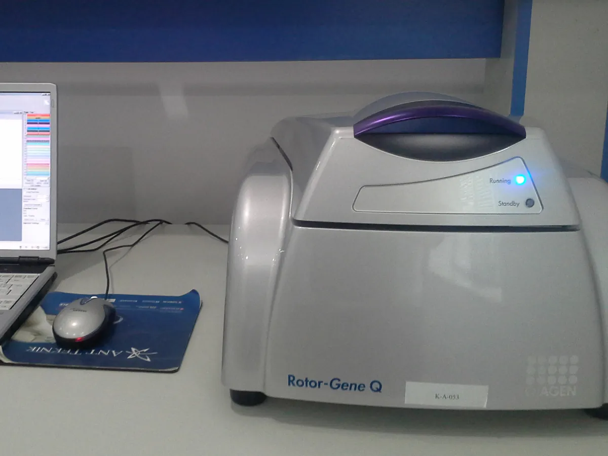
A thermal cycler with a built-in fluorescence reader. It copies a chosen DNA sequence cycle by cycle and measures the fluorescence in every well after each cycle, so the starting amount of DNA can be calculated from a standard curve.
- Biotechnology
- Biopharmaceutical
- Cell and gene therapy
- Academic research
- Agricultural biotech
- Food authenticity
How qPCR works
In each cycle the reaction is heated to separate the DNA strands (denaturation, about 95 °C), cooled so primers bind (annealing, about 55–65 °C) and held so the polymerase extends them (often combined with annealing at 60 °C). Product doubles each cycle at 100% efficiency. Fluorescence from an intercalating dye (such as SYBR Green) or a hydrolysis probe rises in proportion to product. Cq falls by about 3.32 cycles for each ten-fold increase in starting copies, so a plot of Cq against log quantity is a straight line used for quantification.
The path of a sample
- 01
Set up
Master mix, primers, probe, standards, controls and samples are pipetted into a plate, which is sealed with optical film.
- 02
Activate
A hot-start step (typically 95 °C for 2–10 min) activates the polymerase and fully denatures the DNA.
- 03
Cycle
The block cycles 40–45 times between denaturation and annealing/extension temperatures.
- 04
Read
The optics excite each dye and read emission from every well at the end of each annealing/extension step.
- 05
Analyse
Software sets the baseline and threshold, finds Cq for each well and reads quantities from the standard curve.
- 06
Confirm
Optional melt curve (for dye assays) and review of controls, efficiency and replicate agreement before results are reported.
Main parts and what they do
Thermal block (Peltier)
Heats and cools a 96- or 384-well block quickly and evenly; may support a temperature gradient for optimising annealing.
Heated lid
Presses the sealed plate onto the block and stays at about 105 °C so liquid does not condense on the seal.
Excitation source
LEDs or a halogen/tungsten lamp with excitation filters for each dye channel.
Emission filters and detector
A CCD/CMOS camera or photodiodes read the emitted light in each colour channel.
Optical calibration files
Background, uniformity and pure-dye spectra stored on the instrument and used to correct and separate the raw signals.
Software
Controls the run, applies calibrations, calculates Cq, standard curves and melt curves, and keeps an audit trail.
Terms every analyst should know
Quantification cycle (Cq / Ct)
The cycle at which fluorescence crosses the threshold. Lower Cq means more starting template. Replicate Cq values typically agree within about 0.3–0.5 cycles.
Amplification efficiency
E = 10^(−1/slope) − 1 from the standard curve. A slope of −3.32 means 100% efficiency; 90–110% (slope about −3.6 to −3.1) is a common acceptance range.
Passive reference dye
A dye such as ROX that does not take part in the reaction. Dividing the reporter signal by it corrects for small well-to-well differences in volume and optics.
Hydrolysis probe
A short labelled oligonucleotide (for example a TaqMan probe) with a reporter and a quencher. The polymerase cuts it during extension, separating reporter from quencher so fluorescence rises.
Melt curve
After cycling, the product is slowly heated and fluorescence of an intercalating dye drops as strands separate. One sharp peak indicates a single specific product.
Controls
No-template controls (NTC) show contamination; a negative extraction control and a spiked positive control show the extraction and amplification worked.
Keep every qPCR in control: usage logs, calibration and maintenance schedules, and automatic blocking of overdue instruments.