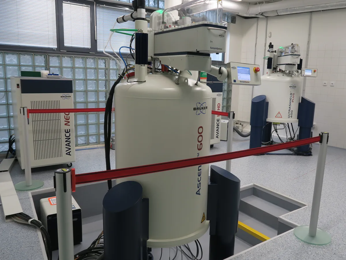
Nuclear magnetic resonance (NMR) spectrometer
Places a sample in a strong magnetic field and measures how atomic nuclei such as ¹H and ¹³C absorb radio waves. Gives detailed structural information and, with qNMR, absolute purity without a reference standard of the analyte.
- Pharmaceutical
- Biotech
- Chemicals
- Academia & research
- Food (authenticity)
- Petrochemical
How NMR works
Nuclei with a spin (e.g. ¹H) align with or against a strong magnetic field B₀, giving two energy levels. A short radio-frequency pulse at the resonance (Larmor) frequency tips the nuclei; as they relax back they emit a signal (free induction decay, FID). A Fourier transform turns the FID into a spectrum. Electrons around each nucleus shield it slightly, so nuclei in different environments resonate at slightly different frequencies, reported as the chemical shift in ppm.
The path of a sample
- 01
Sample in a deuterated solvent
A few milligrams are dissolved in a deuterated solvent (e.g. CDCl₃, DMSO-d₆, D₂O) in a precision NMR tube.
- 02
Lock and shim
The instrument locks onto the deuterium signal to keep the field stable and shims the field to make it uniform across the sample.
- 03
Tune and match
The probe is tuned to the nucleus being observed for maximum sensitivity.
- 04
RF pulse
A short radio-frequency pulse excites the nuclei.
- 05
FID acquisition
The receiver records the decaying signal as the nuclei relax; many scans are added to improve signal-to-noise.
- 06
Processing
Fourier transform, phase and baseline correction, referencing (e.g. TMS at 0 ppm) and integration give the final spectrum.
Main parts and what they do
Superconducting magnet
Generates a very strong, stable field (typically 7–14 T for 300–600 MHz); cooled by liquid helium inside a liquid-nitrogen jacket. Benchtop units use permanent magnets.
Probe
Holds the sample tube in the field centre and contains the coils that transmit RF pulses and receive the signal.
Shim coils
Small adjustable coils that make the field uniform across the sample for sharp lines.
RF console (transmitter and receiver)
Generates the pulses and amplifies and digitises the weak NMR signal.
Lock system
Monitors the deuterium signal from the solvent and corrects field drift.
Sample changer
Automates loading of tubes for unattended runs.
Workstation and software
Controls experiments, processes FIDs and stores data with an audit trail.
Terms every analyst should know
Chemical shift (δ, ppm)
Signal position relative to a reference such as tetramethylsilane (TMS). It is independent of field strength, so spectra from different instruments compare directly.
Integration
The area of a ¹H signal is proportional to the number of protons it represents, e.g. a CH₃ gives three times the area of a CH.
Spin–spin coupling
Neighbouring nuclei split each other's signals (doublet, triplet…). The splitting (J, in Hz) shows which atoms are connected.
Relaxation (T1)
The time nuclei take to return to equilibrium. For qNMR the delay between scans must be long enough (commonly at least 5 × the longest T1) for full relaxation.
Field strength
Quoted as ¹H frequency (e.g. 400 or 600 MHz). Higher fields spread signals further apart and improve sensitivity.
Quantitative NMR (qNMR)
Purity = (Ix/Istd) × (Nstd/Nx) × (Mx/Mstd) × (mstd/mx) × Pstd, using a certified internal standard such as maleic acid or dimethyl sulfone.
2D NMR
COSY (H–H), HSQC (direct C–H) and HMBC (long-range C–H) experiments map out the carbon skeleton for structure elucidation.
Keep every NMR in control: usage logs, calibration and maintenance schedules, and automatic blocking of overdue instruments.