Fluorescence Spectrophotometer (Spectrofluorometer)
Shines light of one wavelength on a sample and measures the longer-wavelength light the molecules give back (fluorescence). Very sensitive and selective for fluorescent compounds.
- Pharmaceutical
- Biotech
- Clinical
- Environmental
- Food & beverage
- Research
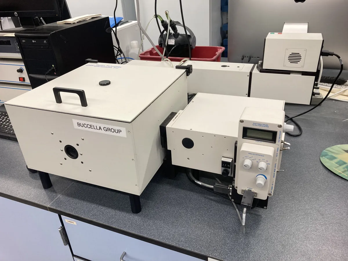
How Fluorimeter works
A molecule absorbs a photon and is raised to an excited electronic state. It quickly loses some energy by vibration, then returns to the ground state by emitting a photon of lower energy (longer wavelength). At low concentrations the emitted intensity is proportional to concentration: F = k × I₀ × Φ × ε × b × c.
The path of a sample
- 01
Light source
A xenon arc or flash lamp gives a continuous spectrum from UV to visible.
- 02
Excitation monochromator
Selects the excitation wavelength (λex) that the analyte absorbs.
- 03
Sample cell
A four-side-polished quartz cuvette holds the sample; fluorescence comes out in all directions.
- 04
Emission monochromator
Placed at 90° to the excitation beam to avoid the transmitted light; selects the emission wavelength (λem).
- 05
Detector
A photomultiplier tube measures the weak emitted light; a reference detector corrects for lamp fluctuation.
Main parts and what they do
Xenon lamp
Broadband continuous light source; flash lamps last longer and reduce photobleaching.
Excitation monochromator
Grating and slits that select the excitation wavelength and bandwidth.
Sample compartment
Holds the quartz cuvette, often with a temperature-controlled (Peltier) holder.
Emission monochromator
Grating and slits at 90° that select the emission wavelength.
Photomultiplier tube (PMT)
Highly sensitive detector; its voltage (gain) sets sensitivity.
Reference detector
Monitors lamp intensity so the ratio signal is stable.
Filters and polarisers (optional)
Remove second-order light or measure fluorescence anisotropy.
Software
Scans spectra, measures intensity, builds calibration curves and keeps the audit trail.
Terms every analyst should know
Stokes shift
The difference between the excitation and emission maxima. Emission is always at a longer wavelength than excitation.
Quantum yield (Φ)
The fraction of absorbed photons that are re-emitted as fluorescence. Strong fluorophores have Φ close to 1.
Inner-filter effect
At high absorbance the solution absorbs the excitation light (or its own emission) before it reaches the viewing zone, so the response curves downwards. Keep absorbance low (often below about 0.05–0.1).
Quenching
Loss of fluorescence by collision with other species (oxygen, halide ions, heavy atoms) or by complex formation.
Water Raman band
Inelastic scatter from water at a fixed energy shift (about 3400 cm⁻¹) from the excitation. With 350 nm excitation it appears near 397 nm and is used to test sensitivity.
Excitation and emission spectra
Scanning one monochromator while holding the other fixed gives the excitation or emission spectrum used to choose wavelengths.
Keep every Fluorimeter in control: usage logs, calibration and maintenance schedules, and automatic blocking of overdue instruments.