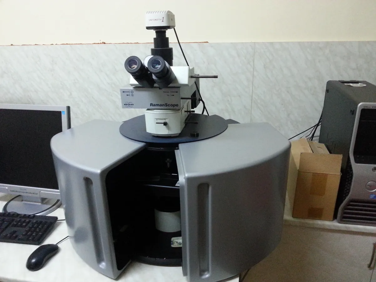
Raman spectrometer
Shines a laser on a sample and measures the tiny fraction of light scattered with a shifted wavelength. Gives a sharp molecular fingerprint through glass and plastic, ideal for raw-material identity and polymorph checks.
- Pharmaceutical
- Biotech
- Forensics & security
- Materials science
- Semiconductors
- Art conservation
How Raman works
When monochromatic laser light hits a molecule, most of it scatters elastically (Rayleigh). A very small fraction exchanges energy with a molecular vibration and scatters at a lower energy (Stokes) or higher energy (anti-Stokes). The difference, the Raman shift in cm⁻¹, equals the vibrational frequency. A vibration is Raman-active when it changes the polarisability of the electron cloud, so symmetric, non-polar bonds give strong bands.
The path of a sample
- 01
Laser
A stable, narrow-line laser (often 785 nm) excites the sample.
- 02
Sampling optics
A probe, vial holder or microscope objective focuses the laser and collects the scattered light.
- 03
Rayleigh filter
A notch or edge filter blocks the intense light at the laser wavelength.
- 04
Spectrograph
A grating (dispersive) or interferometer (FT-Raman) separates the Raman-scattered light by wavelength.
- 05
Detector
A cooled CCD (dispersive) or InGaAs/Ge detector (FT-Raman at 1064 nm) records the spectrum.
- 06
Library comparison
Software corrects the baseline and compares the spectrum with reference spectra to give a pass/fail identity.
Main parts and what they do
Laser
Excitation source; 785 nm diode lasers are most common in pharma, 1064 nm Nd:YAG for fluorescent samples, 532 nm for high sensitivity on non-fluorescent samples.
Probe or sampling accessory
Point-and-shoot nose cone, vial holder, immersion probe or microscope objective that delivers the laser and collects the scatter.
Rayleigh (notch / edge) filter
Blocks the laser line, which is about a million times stronger than the Raman signal.
Spectrograph or interferometer
Separates scattered light by wavelength (dispersive grating or FT interferometer).
Detector
Thermoelectrically cooled CCD for dispersive systems; liquid-nitrogen-cooled Ge or InGaAs for FT-Raman.
Internal calibration standard
Built-in reference (often polystyrene or a neon lamp) used for automatic wavenumber checks.
Software and library
Acquires spectra, runs the identification method and keeps an audit trail.
Terms every analyst should know
Raman shift
The energy difference between the laser and the scattered light, in cm⁻¹. It is independent of the laser used, so spectra from 785 nm and 1064 nm systems line up.
Stokes and anti-Stokes
Stokes lines (lower energy) are much stronger at room temperature and are the ones normally recorded.
Polarisability
Raman activity needs a change in polarisability; IR activity needs a change in dipole moment. This is why the two techniques complement each other.
Fluorescence background
Fluorescence produces a broad, rising baseline that can hide Raman bands. Longer wavelength lasers (785 or 1064 nm) reduce it.
Library identification
The test spectrum is compared with reference spectra using a correlation (hit quality index) or a statistical p-value approach against a threshold set during validation.
Polymorph sensitivity
Low-wavenumber lattice modes and small band shifts reflect crystal packing, so Raman can tell polymorphs apart.
Keep every Raman in control: usage logs, calibration and maintenance schedules, and automatic blocking of overdue instruments.