Gas Chromatography–Mass Spectrometry (GC-MS)
Couples a gas chromatograph to a mass spectrometer so each volatile compound is separated and then identified from its fragmentation pattern. Used for unknown volatile impurities, nitrosamines, residual solvent confirmation and extractables.
- Pharmaceutical
- Environmental
- Forensic and toxicology
- Food & flavour
- Petrochemical
- Packaging
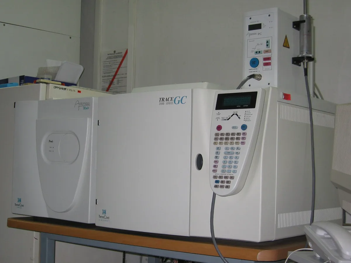
How GC-MS works
Volatile compounds are separated on a capillary GC column and enter the ion source, where 70 eV electrons ionise and fragment them. A quadrupole (most commonly) filters ions by m/z, either scanning a mass range to record full spectra or monitoring selected ions for sensitivity. The total ion chromatogram shows every compound; extracted ion chromatograms show only selected masses.
The path of a sample
- 01
Injection
The sample is vaporised in a heated split / splitless inlet, or introduced by headspace or thermal desorption.
- 02
GC separation
Compounds separate on a low-bleed capillary column (often 30 m × 0.25 mm) under a temperature programme.
- 03
Transfer line
A heated line carries the column outlet into the MS vacuum without condensation.
- 04
EI ion source
A filament emits electrons at 70 eV that ionise and fragment the molecules.
- 05
Quadrupole analyser
Filters ions by m/z, either scanning (e.g. m/z 35–500) or in SIM mode.
- 06
Detector and library search
An electron multiplier records ions; spectra are compared with a reference library to suggest an identity.
Main parts and what they do
Gas chromatograph with inlet and oven
Separates the sample; the inlet liner and septum must be clean and inert.
Low-bleed MS capillary column
Separates compounds with minimal stationary-phase bleed into the MS.
Heated transfer line
Delivers the column effluent into the ion source at a controlled temperature.
Ion source (EI, optionally CI)
Filament and repeller ionise and push ions toward the analyser.
Quadrupole mass filter
Four rods with RF and DC voltages that pass only ions of a chosen m/z.
Electron multiplier detector
Amplifies the ion signal; its voltage rises as it ages.
Vacuum system
Rough pump plus turbomolecular (or diffusion) pump maintain high vacuum.
Calibrant vial (PFTBA)
Supplies the reference compound for tuning and mass-axis calibration.
Data system and spectral library
Controls acquisition, processes data and searches spectra against reference libraries.
Terms every analyst should know
Electron ionisation (EI)
A hard ionisation method at a standard 70 eV that gives reproducible fragment patterns, the basis of library searching.
Total ion chromatogram (TIC)
The sum of all ion currents at each time point; it looks like an ordinary chromatogram.
Selected ion monitoring (SIM)
The quadrupole monitors only a few chosen m/z values, giving much higher sensitivity than full scan.
Base peak and molecular ion
The base peak is the most intense ion (set to 100%); the molecular ion (M⁺•) shows the molecular weight if it survives.
Library match
A similarity score between the measured and library spectra. A good match supports, but does not prove, identity; confirm with a reference standard.
Autotune
An automatic routine that uses a reference compound (usually PFTBA) to set lens voltages and calibrate the mass axis.
Keep every GC-MS in control: usage logs, calibration and maintenance schedules, and automatic blocking of overdue instruments.