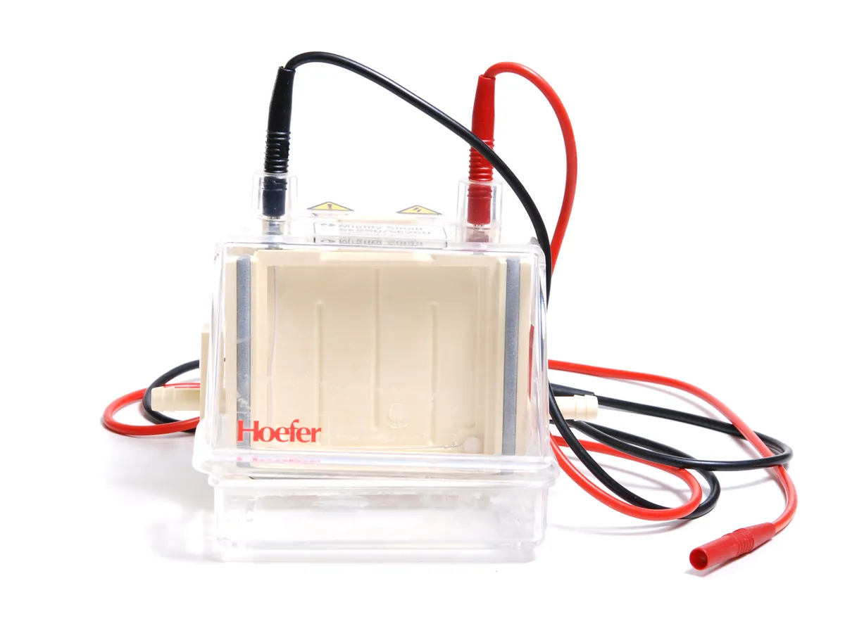
Gel electrophoresis system
A tank with buffer and electrodes, a gel and a DC power supply. Charged molecules such as proteins or DNA move through the gel in the electric field and separate by size, then are stained and imaged.
- Biotechnology
- Biopharmaceutical
- Academic research
- Diagnostics development
- Food (protein profiling)
- Vaccines
How Gel electrophoresis works
A charged molecule in an electric field moves towards the electrode of opposite charge. Its speed depends on charge, size and shape, and on the field strength (voltage per cm). The gel acts as a sieve, slowing larger molecules more than smaller ones. With SDS (proteins) or the uniform charge of DNA, mobility depends mainly on size, so band position is roughly linear with the logarithm of molecular weight over the useful range of the gel.
The path of a sample
- 01
Prepare gel and buffer
Cast or buy a gel of suitable percentage; fill the tank with running buffer.
- 02
Prepare samples
Mix samples with loading buffer (with SDS and reducing agent for reducing SDS-PAGE) and heat as the method says.
- 03
Load
Load samples, a molecular weight marker and any reference standard into the wells.
- 04
Run
Apply constant voltage (or current) until the dye front nears the bottom of the gel.
- 05
Stain and image
Stain the gel, destain if needed and capture an image with a gel documentation system.
- 06
Evaluate
Compare band pattern with the reference; estimate size from the marker and, if required, quantify bands by densitometry.
Main parts and what they do
Gel tank
Holds buffer and gel; vertical for PAGE, horizontal for agarose. Lid with interlocked electrical connectors.
Electrodes
Platinum wires that carry current into the buffer.
Gel cassette / casting tray and combs
Hold the gel and form sample wells.
DC power supply
Provides constant voltage, current or power with timers, and shuts off on open circuit, leaks or overload.
Gel imager
White-light, UV or blue-light transilluminator and camera to document stained gels.
Cooling (optional)
Cooling core or circulator to remove heat for long or high-voltage runs.
Terms every analyst should know
SDS-PAGE
Sodium dodecyl sulphate coats proteins with negative charge in proportion to their length, so proteins separate by size. A reducing agent (DTT or β-mercaptoethanol) breaks disulphide bonds.
Gel percentage
Higher acrylamide or agarose percentage gives smaller pores and better separation of small molecules. Gradient gels cover a wider range.
Ohm's law
V = I × R. At constant voltage, current falls as buffer ions are used up and resistance rises. Power (W = V × I) is turned into heat.
Joule heating
Heat from the current. Too much causes band smiling, distortion and gel damage, so voltage limits and cooling matter.
Molecular weight marker
A ladder of proteins or DNA fragments of known size run alongside samples to estimate size.
Densitometry
Measuring band intensity from the gel image to estimate relative amounts, for example the per cent of a main band.
Keep every Gel electrophoresis in control: usage logs, calibration and maintenance schedules, and automatic blocking of overdue instruments.