Colony counter
Helps the analyst count colonies on agar plates and membranes accurately, either by a lit magnifier with a counting pen (manual) or by a camera and image analysis software (automated).
- Pharmaceutical
- Biotech
- Food & beverage
- Water testing
- Cosmetics
- Research laboratories
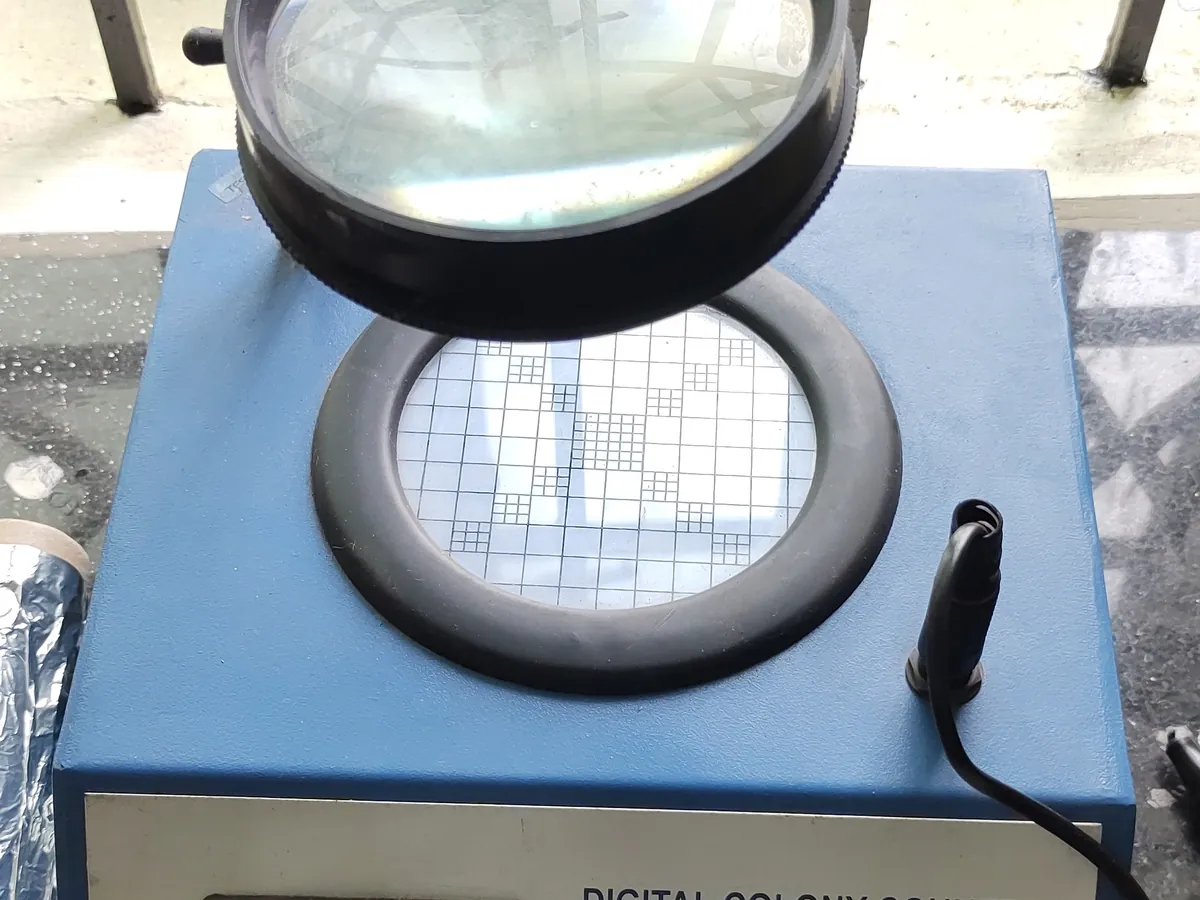
How Colony counter works
Colonies are made more visible by contrast lighting and magnification. In manual counters, each colony is marked by touching it with a pen connected to an electronic counter. In automated counters, a camera image is processed by software that separates colonies from the background by contrast and size, then counts them.
The path of a sample
- 01
Place plate
The plate or membrane is placed on the lit stage, lid removed or inverted as per the SOP.
- 02
Illuminate
Back-light or dark-field lighting increases contrast between colonies and agar.
- 03
Magnify / capture
A lens magnifies the plate (manual) or a camera captures an image (automated).
- 04
Mark / detect
The analyst marks each colony, or software detects colonies by contrast, size and shape.
- 05
Review & record
The analyst checks the count, edits software marks if needed and records the result.
Main parts and what they do
Viewing stage
Holds the plate over a light source; often with a counting grid.
Light source
Back-light or dark-field ring light to increase colony contrast.
Magnifier lens
Typically 1.5–3× magnification for manual counting.
Counting pen or pressure pad
Registers a count each time a colony is touched and marks it to prevent double counting.
Camera and software (automated)
Captures the plate image, detects colonies and stores the result with the image.
Display / counter
Shows the running count; automated systems export results to a LIMS.
Terms every analyst should know
Countable range
The range of colonies per plate that gives a reliable count, for example not more than 250 cfu for bacteria and 50 cfu for fungi on a 9 cm plate (USP <61> approach).
Dark-field vs bright-field
Dark-field lighting makes colonies glow against a dark background and suits clear agar; bright-field back-lighting suits coloured media.
Detection threshold
In automated counters, the minimum colony size and contrast counted. Too low counts debris; too high misses small colonies.
Data integrity
Automated counter images and results are GMP records: they need user log-in, audit trail and secure backup.
Keep every Colony counter in control: usage logs, calibration and maintenance schedules, and automatic blocking of overdue instruments.