Particle & solid-state
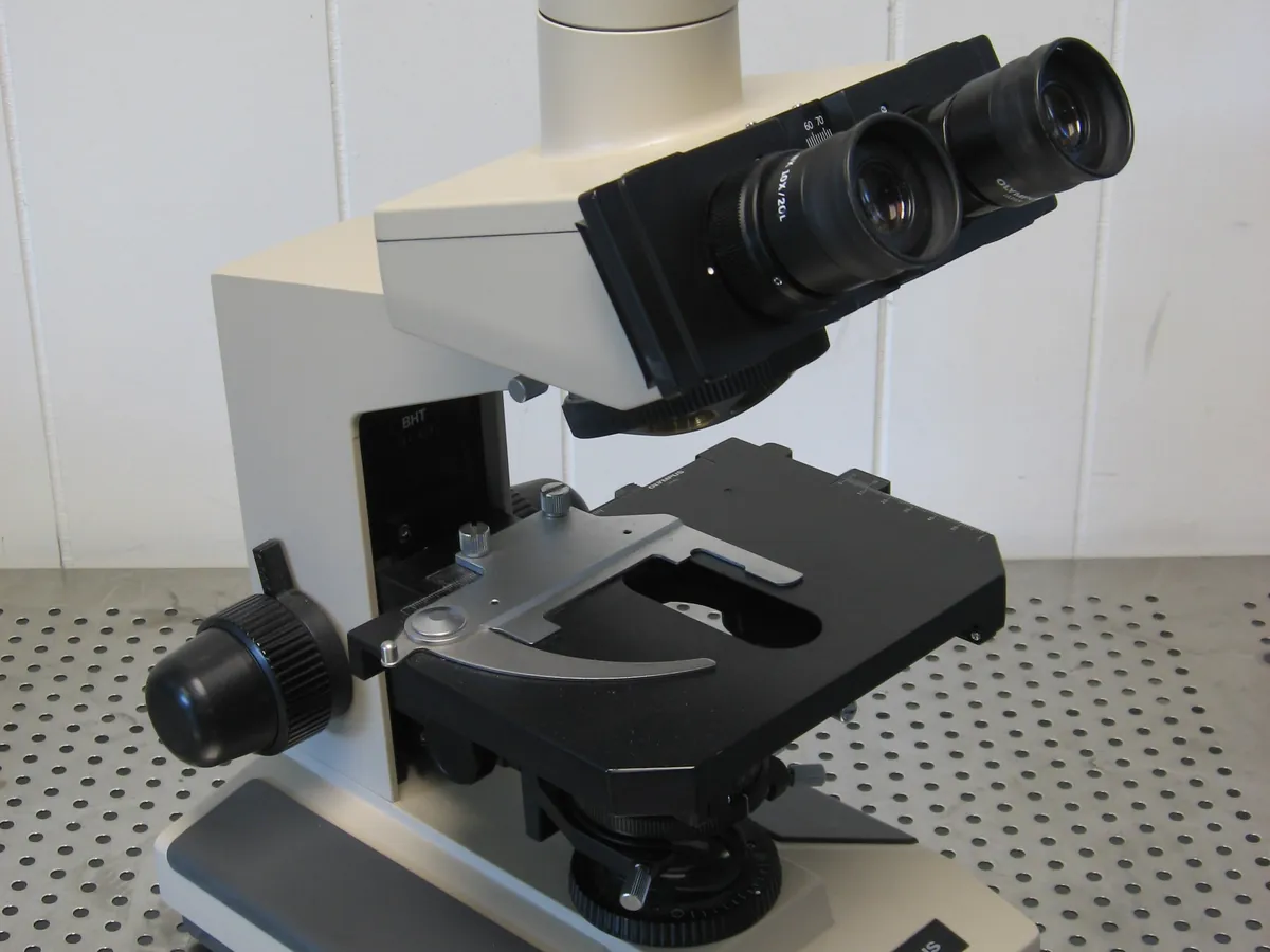
Optical (Light) Microscope
Magnifies particles so their size, shape and crystal habit can be seen and measured with a calibrated eyepiece graticule or camera. Used for particle size, morphology and identification checks.
- Pharmaceutical
- Microbiology
- Materials
- Food
- Forensics
- Education
Follow your site SOP
This is a typical operating sequence for learning. Your instrument model and approved SOP take precedence.
STAGE 1
Setup
- 1Check the microscope is clean and its graticule calibration is current.
- 2Switch on the lamp at low intensity and set Köhler illumination as per the manual.
- 3Calibrate the graticule with a stage micrometer for each objective used, if not already recorded.
STAGE 2
Sample preparation
- 1Disperse a small amount of powder in a mounting fluid in which it does not dissolve (e.g. mineral oil).
- 2Place a drop on a clean slide and lower a cover slip at an angle to avoid air bubbles.
- Prepare potent or toxic samples in a containment hood.
STAGE 3
Observation and measurement
- 1Start with the low-power objective to find the field, then move to higher power.
- 2Scan several fields in a set pattern so you do not choose particles subjectively.
- 3Measure the required number of particles and record their size and shape.
- 4Switch on the crossed polarisers to check for birefringence if required.
- 5Capture representative images for the record.
- Never lower a high-power objective towards the slide while looking through the eyepiece; you may crush the slide and damage the lens.
STAGE 4
After use
- 1Remove the slide; clean oil from the immersion objective with lens tissue
- 2Lower the lamp intensity and switch off
- 3Cover the microscope
In LimsPulse · Calibration Management
Keep every Optical microscope in control: usage logs, calibration and maintenance schedules, and automatic blocking of overdue instruments.