Optical (Light) Microscope
Magnifies particles so their size, shape and crystal habit can be seen and measured with a calibrated eyepiece graticule or camera. Used for particle size, morphology and identification checks.
- Pharmaceutical
- Microbiology
- Materials
- Food
- Forensics
- Education
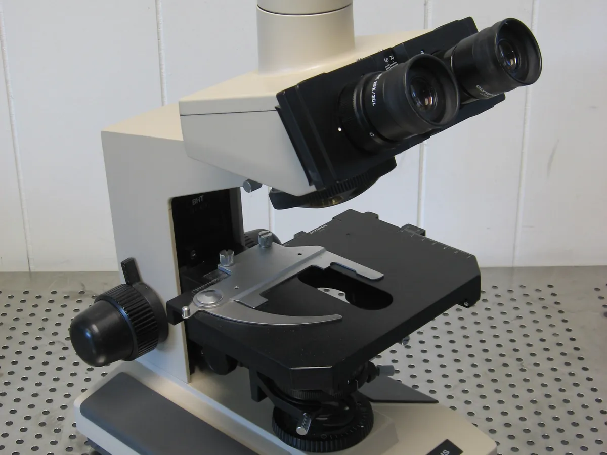
How Optical microscope works
Light passes through (or reflects from) the sample and is focused by the objective lens to form a magnified image, which the eyepiece magnifies further. Total magnification is objective × eyepiece. Resolution is limited by the wavelength of light and the numerical aperture of the objective.
The path of a sample
- 01
Illumination
A lamp and condenser send an even cone of light through the slide.
- 02
Sample
Particles are dispersed in a suitable mounting fluid on a glass slide with a cover slip.
- 03
Objective
Forms a magnified real image of the particles (e.g. 4×, 10×, 40×, 100×).
- 04
Eyepiece / camera
Magnifies the image further (usually 10×) or projects it onto a camera.
- 05
Measurement
Particles are sized against a graticule calibrated with a stage micrometer.
Main parts and what they do
Light source and condenser
Provide bright, even illumination focused on the sample.
Stage
Holds the slide and moves it in X and Y for scanning.
Objective lenses
Main magnifying lenses on a rotating nosepiece.
Eyepieces with graticule
Magnify the image; the graticule scale is used for sizing.
Polariser and analyser
Filters used for polarised light microscopy.
Camera and image software
Capture images and measure particles with a calibrated scale.
Terms every analyst should know
Magnification
Objective × eyepiece; for example 40× × 10× = 400×.
Resolution
The smallest distance at which two points can be seen as separate; about 0.2 µm at best with visible light and oil immersion.
Stage micrometer
A slide with an accurately ruled scale (e.g. 1 mm in 100 divisions of 10 µm) used to calibrate the eyepiece graticule.
Birefringence
Crystalline particles split polarised light and appear bright against a dark background under crossed polarisers.
Feret and Martin diameters
Common ways to define size of irregular particles: Feret is the distance between parallel tangents; Martin is the length of the line that splits the projected area in half.
Keep every Optical microscope in control: usage logs, calibration and maintenance schedules, and automatic blocking of overdue instruments.